Author: Catherine Goh

Ribo-depletion in RNA-Seq – Which ribosomal RNA depletion method works best?

Ribo-depletion in RNA-Seq – Which ribosomal RNA depletion method works best?

Summary: This blogpost is focussed on ribosomal RNA (rRNA) depletion methods frequently applied to improve and economize RNA-Seq experiments.

The Rise of RNA-Seq

RNA-Seq Overtakes Microarrays

The use of Next-Generation RNA Sequencing (RNA-Seq) has recently overtaken that of DNA-based microarrays to detect and quantify changes in gene expression.

Why? RNA-Seq can detect novel coding and non-coding genes, splice isoforms, single nucleotide variants and gene fusions. Its broader dynamic range also enables sensitive detection of low abundance transcripts.

RNA-Seq vs Microarray

Also, technological advancements in single cell isolation, ribosome profiling and pulse-labelling techniques can now be multiplexed with RNA-Seq to widen the scope of scientific interrogation. Now, one can study the transcriptome, translotome and epitranscriptome with added spatial and temporal resolution. Studies of RNA structure and  RNA-protein/nucleic acid interactions with the use of nuclease digestion and biochemical pulldown approaches have also increasingly employed RNA-Seq. This excellent review describes all these latest advances.

A Major Challenge in RNA-Seq

A major limitation encountered in RNA-Seq however is the massive abundance of ribosomal RNA (rRNA) that can occupy up to 90% of RNA-Seq reads.  This necessitates additional steps for ribo-depletion or rRNA depletion to economize an RNA-Seq experiment.

Ribo-Depletion Methods

1) Poly-A selection

The most common method of rRNA depletion is poly-A selection, which relies on the use of oligo (dT) primers attached to a solid support (e.g. magnetic beads) to isolate protein-coding polyadenylated RNA transcripts. The main disadvantage though is one misses out on non-polyadenylated transcripts which include microRNAs, small nucleolar RNAs (snoRNAs), some long non-coding RNAs (lncRNA), and even some protein-coding RNAs (histones) which lack polyA tails. As a result, one fails to capture biologically relevant insights on these RNAs which make up a substantial proportion of the transcriptome.

Poly-A Selection - Advantages and Disadvantages

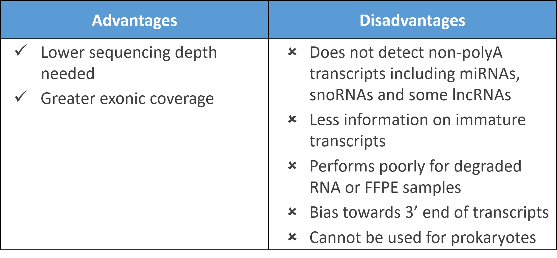
Curiously, polyadenylated transcripts are more abundant in eukaryotes as opposed to prokaryotes with both groups using polyadenylation in entirely different ways! Hence, polyA selection cannot be applied for sequencing of bacteria and archaebacteria, excluding its use in metagenomic RNA-Seq.

Poly-A selection also relies on transcripts being largely intact and tend to over-represent 3′ regions of transcripts. Studies comparing physical rRNA depletion methods and polyA selection show polyA selection did not work well for degraded RNA samples. A lower number of reads were also obtained with formalin-fixed paraffin-embedded (FFPE) tissues though analysis of fresh frozen tissues was not compromised.

Despite this, polyA selection still provides greater exonic coverage than physical rRNA depletion which tend to produce more intronic reads.  Further, a lower sequencing depth is typically needed for polyA selection, making it a respectable choice if one is focused only on protein-coding genes.

2) Physical Ribosomal RNA (rRNA) removal

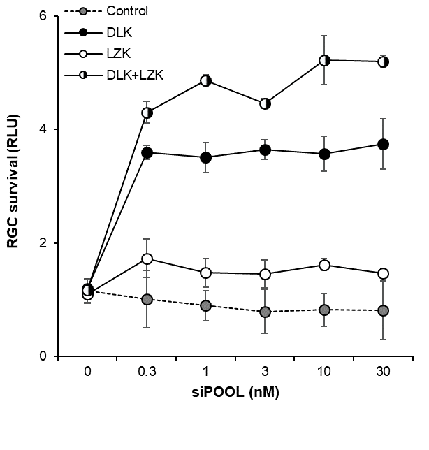
Ribosomal rRNA can also be removed by hybridization to complementary biotinylated oligo probes, followed by extraction with streptavidin-coated magnetic beads. riboPOOLs developed by siTOOLs Biotech efficiently removes rRNA through this route, with a workflow similar to Ribo-Zero from Illumina.

Physical rRNA removal workflow
Workflow for rRNA removal with biotinylated probes and streptavidin-coated magnetic beads

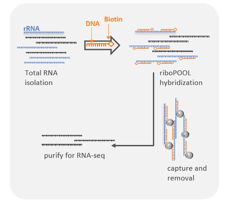
Compared to polyA selection methods, rRNA removal enables detection of non-polyadenylated transcripts and small RNAs.  Comparisons between differential gene expression detected with both methods were typically well-correlated. The rRNA removal method however could detect both long and short transcripts showing less of a 3′ bias than polyA selection.

Physical rRNA removal also performs better for degraded and FFPE samples, and can also be applied for metagenomic samples that contain microbes. The Pan-Prokaryote riboPOOL by siTOOLs for example, functions effectively to remove rRNA from a diverse range of prokaryotic species, and can be used in combination with human and mouse/rat riboPOOLs to deplete rRNA from complex samples containing multiple species.

Physical rRNA Removal - Advantages and Disadvantages

By using targeted probes, one can also flexibly deplete abundant RNAs that take up expensive RNA-Seq reads. For example, globin, found in high amounts in RNA isolated from blood samples, can be efficiently depleted by globin mRNA-specific probes.

Ribosomal RNA can also be removed by selective degradation where enzyme RNase H is used to specifically degrade DNA-RNA hybrids formed between DNA probes and complementary rRNA (e.g. NEBNext rRNA depletion kit by New England Biolabs). This method was reported to produce consistent results, working as well on degraded samples though there was a bias against detecting transcripts of shorter length compared to Ribo-Zero.

3) Targeted amplification

An alternative method to deplete rRNA involves the use of  “not so random” hexamer/heptamer primers with a decreased affinity for rRNA during first strand cDNA synthesis. This is employed by the Ovation RNA-Seq kits from NuGen. Though the kit can be used to detect non-polyA RNAs and can be applied to prokaryotes, the additional incorporation of oligo(dT) still contributes to a bias towards  detecting 3′ regions.

A recent ribosome profiling study comparing library preparation methods reported fewer reads obtained and greater intronic reads for Nugen kits compared to polyA-selection methods. As Nugen also incorporated an RNase-mediated degradation of unwanted transcripts during final library construction steps, this indicates targeted amplification alone cannot totally remove rRNA. The method does however work with low input amounts and degraded samples.

 Targeted Amplification with not so random primers - Advantages and Disadvantages

So which ribo-depletion method works best?

And the answer as always? It depends. Depending on the ribo-depletion method chosen in RNA-Seq library preparation, some differences in genes detected and their expression levels will certainly be observed.

Poly-A selection might be the most efficient method when only focussed on protein-coding genes, but one loses significant information on non-polyadenylated RNAs and immature transcripts. In instances such as microbial sequencing or in sequencing degraded or FFPE samples, poly-A selection cannot even be applied or may perform poorly.

Physical rRNA removal offers the advantage of retrieving more transcriptomic information but comes at a cost of greater intronic/intergenic reads that necessitates a greater sequencing depth. However, it offers greater flexibility and better performance in sequencing of challenging sample types.

Targeted amplification with “not so random” primers though effective for low input material, comes also at a cost of greater sequencing depth required.

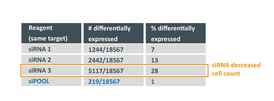
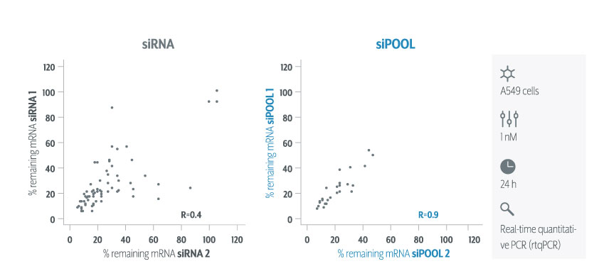
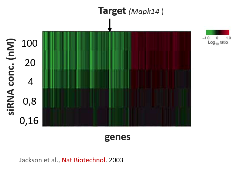
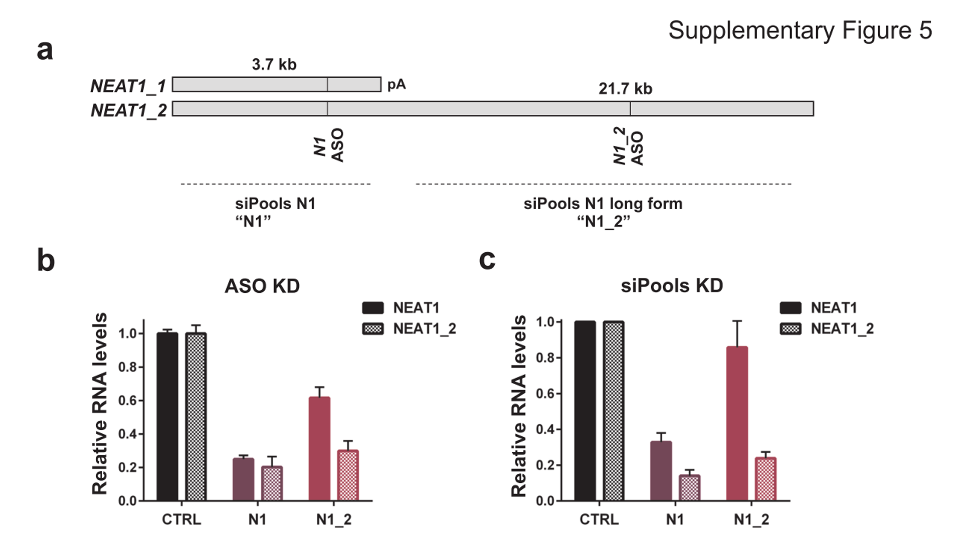
All methods are subject to some extent of non-specificity and detection bias. Further variability can also arise from different methods of sequence alignment in RNA-Seq data analysis. It is therefore always advisable to validate sequencing data obtained by real-time quantitative PCR (rtqPCR) or other methods.

References:

1. Song, Y., Milon, B., Ott, S., Zhao, X., Sadzewicz, L., Shetty, A., Boger, E. T., Tallon, L. J., Morell, R. J., Mahurkar, A., and Hertzano, R. (2018) A comparative analysis of library prep approaches for sequencing low input translatome samples. BMC Genomics. 19, 696
2. O’Neil, D., Glowatz, H., and Schlumpberger, M. (2013) Ribosomal RNA Depletion for Efficient Use of RNA-Seq Capacity. in Current Protocols in Molecular Biology, p. 4.19.1-4.19.8, John Wiley & Sons, Inc., Hoboken, NJ, USA, 103, 4.19.1-4.19.8
3. Stark, R., Grzelak, M., and Hadfield, J. (2019) RNA sequencing: the teenage years. Nat. Rev. Genet. 10.1038/s41576-019-0150-2
4. Cui, P., Lin, Q., Ding, F., Xin, C., Gong, W., Zhang, L., Geng, J., Zhang, B., Yu, X., Yang, J., Hu, S., and Yu, J. (2010) A comparison between ribo-minus RNA-sequencing and polyA-selected RNA-sequencing. Genomics. 96, 259–265
5. Zhao, S., Zhang, Y., Gamini, R., Zhang, B., and von Schack, D. (2018) Evaluation of two main RNA-seq approaches for gene quantification in clinical RNA sequencing: polyA+ selection versus rRNA depletion. Sci. Rep. 8, 4781
6. Herbert, Z. T., Kershner, J. P., Butty, V. L., Thimmapuram, J., Choudhari, S., Alekseyev, Y. O., Fan, J., Podnar, J. W., Wilcox, E., Gipson, J., Gillaspy, A., Jepsen, K., BonDurant, S. S., Morris, K., Berkeley, M., LeClerc, A., Simpson, S. D., Sommerville, G., Grimmett, L., Adams, M., and Levine, S. S. (2018) Cross-site comparison of ribosomal depletion kits for Illumina RNAseq library construction. BMC Genomics. 19, 199

Featured Image is an artist’s rendition of a ribosome. Credit: C. BICKLE/SCIENCE

Performing target validation well

Performing target validation well

Summary

This blogpost describes issues encountered in target validation and how to safeguard against poor reproducibility in RNAi experiments.

The importance of target validation

More than half of all clinical trials fail from a lack of drug efficacy. One of the major reasons for this is inadequate target validation.

Target validation involves verifying whether a target (protein/nucleic acid) merits the development of a drug (small molecule/biologic) for therapeutic application.

Failing to adequately validate a target can burden a pharma with roughly 800 million to 1.4 billion in drug development costs. Impact is not only monetary as large site closures  often result as companies struggle to save costs and a reduced production effort deprives patients of new medicines.

Performing target validation well

Special attention should therefore be given to performing target validation techniques well.

target validation techniques
Overview of target validation techniques (Lindsay, Nat Review Drug Discovery, 2013)

Many of these techniques involve inhibiting target expression to establish its relevance in a cellular or animal disease model. This can be performed with chemical probes, RNA interference (RNAi), genetic knock-outs, and even targeted protein degradation.

The reproducibility of these techniques however has been an issue of concern for drug developers. Less than half of all findings from peer-reviewed scientific publications was reported to be successfully reproduced.

Dismal rates of reproducibility from several pharma-led cancer-focused studies ranged from 11% (Amgen) to 25% (Bayer). A review by William Kaelin Jr sums up the common pitfalls of preclinical cancer target validation. One of his key points:

Cellular phenotypes caused by a chemical or genetic perturbant should be considered to be off-target until proved otherwise, especially when the phenotypes were detected in a down assay and therefore could reflect a nonspecific loss of cellular fitness. It is only by performing rescue experiments that one can formally address whether the effects of a perturbant are on-target.

The comment highlights the issue of reagent non-specificity as a notable contribution towards poor reproducibility.

Certainly, for RNAi the wide-spread off-target effects of siRNAs has been observed in numerous publications. The mechanism being well-established to be based on microRNA-like seed-based recognition of non-target genes. The effect dominates over on-target effects in many large RNAi screens, illustrating the depth of the problem.

Reagent non-specificity is not restricted to RNAi. There have been multiple reports of non-specificity for gene editing technique, CRISPR, which can be read about in detail here, here and here. Recent publications continue to shed more light on its potential off-targets as we learn more about this relatively new technique.

Even chemical probes may have multiple targets. It is hence imperative that more than one target validation technique be used to avoid confirmation bias.

Target validation – a story from Pharma

Back in 2013, when siTOOLs was just starting out, a pharma approached us with a target validation problem.

They were obtaining different results with 3 different siRNAs in a cellular proliferation assay. Despite all 3 siRNAs potently downregulating the target gene, they produced different effects on cell viability.

Which siRNA tool to trust?

target validation siRNA vs siPOOL pharma story
Three different siRNAs against the same target were tested in a cell proliferation assay. Despite all 3 siRNAs showing potent target gene silencing, effect on cell proliferation differed greatly.

A whole-transcriptome expression analysis performed for the 3 siRNAs and a siPOOL designed against the same target revealed the reason for the large variability.

target validation expression analysis siRNA vs siPOOL pharma story
How many genes can you affect with an siRNA? Whole transcriptome analysis by microarray was performed and number and % of up and down-regulated genes are shown over total number of genes assayed (18567).

Despite all siRNA tools affecting the same target, the difference in extent of gene deregulation was astounding. With the greatest number of off-target effects, it was not surprising that siRNA 3 showed an impact on cell proliferation.

In contrast, siPOOLs had 5 to 25X less differentially expressed genes compared to the 3 commercial siRNAs against the same target. An expression analysis carried out for another gene target showed similar results i.e. siPOOLs having far less off-targets.

The target was dropped from development. A great example where failing early is a good thing, though it was not without costs from validating the multiple siRNAs.

The recommended target validation tool

Functioning like a pack of wolves, siPOOLs increase the chances of capturing large and difficult prey, while making full use of group diversity to compensate for individual weakness.

siPOOLs efficiently counter RNAi off-target effects by high complexity pooling of sequence-defined siRNAs. This enables individual siRNAs to be administered at much lower concentrations, below the threshold for stimulating significant off-target gene deregulation. Due to having multiple siRNAs against the same target gene, target gene knock-down is maintained and in fact becomes more efficient.

siRNA vs siPOOL rtqPCR knock-down efficiency
siPOOLs increase targeting efficiency, avoiding knock-down variability. Figure shows rtqPCR quantification of target RNA levels when two siPOOLs vs two siRNAs against 36 genes were tested.

We still recommend using multiple target validation techniques. As a first evaluation however, siPOOLs are quick, easy and most of all, reliable.

Rescue experiments can also be performed with siPOOL-resistant rescue constructs.

Find out more

5 factors to consider in multi-gene targeting RNAi screens

5 factors to consider in multi-gene targeting RNAi screens

Summary: Effective functional genomic screening depends on a variety of factors that need to be simultaneously addressed to obtain meaningful results. A recent Cell Reports paper demonstrates this by taking a holistic approach to siRNA screening with the use of multi-isoform/multi-gene targeting to address redundant paralogs and pathways in cancer cells.

The case for multi-gene targeting

Many RNAi screens use arrayed single gene knockdowns to find genes that play an important role in a biological process. The idea is that a single bullet is enough to take down its target leaving a gaping hole that one cannot fail to notice. In some cases, this is true, and is certainly relied upon by drug developers seeking to create specific mono-target drugs.  However, in complex diseases like cancer, cells have evolved fail-safe mechanisms to make them more resistant to external assaults. A single bullet is simply not enough.

Take for example oncogenic protein RAF or Rapidly Accelerated Fibrosarcoma, a tyrosine kinase effector that is a component of the MAPK signalling pathway (Ras-Raf-MEK-ERK). RAF has three isoforms – ARAF, BRAF and RAF1 (also called CRAF). Studies in mouse embryonic development show they all share some form of functional redundancy as knocking out two isoforms produces more severe effects than knocking out each isoform alone.

Screens that target single genes/isoforms therefore tends to bias results towards genes that have no paralogs or only have single isoforms. This was indeed the reason why classical Ras effectors were not identified in previous screens.

Factors to consider in a multi-gene targeting RNAi screen

Determining gene combinations that make sense

The authors of the study did a focussed siRNA screen on 41 RAS effector nodes represented by 84 genes. Out of the 41 nodes, 25 of them had 2-4 functional paralogs where combinatorial gene silencing was carried out with multiple siRNAs. 5 nodes knocked down multiple members of a protein complex. 5 nodes had siRNAs targeting multiple steps within a pathway. Only 6 nodes silenced single genes (highlighted).

Multi-gene targeting screen design

The only caveat with designing such a screen is the requirement for prior knowledge to perform meaningful gene silencing combinations. In this instance, many of the Ras effector pathways are characterized sufficiently to do this well however in other less studied fields, this could be a challenge. Useful tools that would help in designing gene knockdown combinations would include pathway or phenotype databases such as KEGG, REACTOME or Wikipathways. The Phenovault which siTOOLs Biotech is developing, is yet another potentially useful tool.. more details to come!

Number and types of phenotypes

The authors also highlight how a screen that reads only one phenotype might miss other important gene functions. Many RNAi screens sadly still stick to measuring cell proliferation as their only read-out which is greatly influenced by siRNA off-target effects. Here, 5 different phenotypes were measured (cell size, proliferation, apoptosis, reactive oxygen species [ROS], and viability). It was noted that silencing of Cdc42 had little effect on cell viability yet a prominent effect on ROS levels.

To take this up a notch, analysis was also performed at the single-cell level in cells expressing uniform levels of GFP and co-transfected with GFP siRNA. This allowed authors to correlate phenotypes with levels of gene knockdown, generating dose-response curves. How clever!

A lot more work, but adds to data robustness especially when using single siRNAs that are known to be rather variable.

Heterogeneity of cell lines

Many reports and our own observations attest to the heterogenous response of different cell lines to the same treatment. In cancer especially, the large heterogeneity necessitates the use of multiple cell lines. Not doing so would be failing to account for the large genetic diversity observed in the clinic. The authors screened 92 cell lines derived from lung, pancreas and colorectal tissue.

Despite seeing heterogenous responses to node knockdowns, phenotypic responses could be distinguished into  several groups based on effector engagement.  A major group dependended on RAF through direct binding with KRAS, a second major group worked via RSK p90 S6 kinases to drive RSK-mTOR signalling. And a third minor group was dependent on RalGDS. They went on to focus on the first two major groups, naming them KRAS-type and RSK-type respectively.

Reagents – choosing siRNAs and siRNA concentrations

The authors used previously characterized siRNAs to select for more potent siRNAs. This involved an RNAi sensor reporter-based assay that required the generation of 20,000 clones. The reporter was also shRNA-based. Due to heterogeneity in Dicer-mediated cleavage of shRNA, its uncertain if knockdown potency is accurately reflected when translated to siRNAs (read about the difference between shRNAs and siRNAs).

siRNA off-target effects are concentration-dependent

In any case, its a lot of work to characterize all siRNAs to be used in a screen. Furthermore, off-target effects are not addressed.

The authors stuck to a maximal concentration of 12 nM where 2 nM of siRNA was applied per gene. At 2 nM per siRNA, one still risks deregulating other genes. One of the first papers by Aimee Jackson et al., demonstrated an siRNA targeting MAPK14 deregulated many other genes even at concentrations of 1-4 nM.

An important consideration is to ensure total siRNA concentrations are kept constant. In which case, a negative control siRNA has to match or follow the maximal siRNA concentration used. Using different levels of siRNAs runs the risk of biasing off-target effects towards sequences present at higher concentrations.

To learn what the causes, extent and consequences of siRNA off-target effects are, read siTOOLs Technote 1)

Validating results

As with all scientific hypothesis, it helps to arrive at the same conclusion with different approaches.

The two different effector response subgroups identified also responded differently to small molecules. The KRAS-type lines being more sensitive to EGFR and ERK inhibition while the RSK-type lines more sensitive to inhibitors of PDK1, RSK, MTOR, S6K1 and DNA repair enzymes. This was attributed to the latter’s higher basal metabolic activity manifested in larger investments towards oxidative phosphorylation and mitochondrial ribosome maintenance.

By also projecting signatures obtained from cell lines into patient samples (in The Cancer Genome Atlas, TCGA), the subtypes were also effective at predicting differential sensitivity to multiple drug treatments. This highlights the importance in designing effective drug combinations in cancer.

Interestingly, the authors also performed CRISPR pooled screens in parallel. However, due to the restraints of being only able to knockout 1 gene at a time, smaller effects were seen due to gene redundancy. However, they did go on to use CRISPR as well to mutate key genes to affirm the pathway relationships established.

siPOOLs have been used successfully for multi-gene targeting for up to 4 genes, and potentially more. They also safely address off-target effects by high complexity pooling, enabling each siRNA to be applied at picomolar concentrations. For more articles on multi-gene targeting, read an older blogpost:

Understanding gene networks with combinatorial gene knockdown


Disrupting lncRNA function with siPOOLs (RNAi), antisense oligos and CRISPR

Disrupting lncRNA function with siPOOLs (RNAi), antisense oligos and CRISPR

Summary

This blogpost covers methods used in the disruption of lncRNA function. Specifically focusing on RNA interference (with siPOOLs), antisense oligos, and CRISPR approaches. Challenges faced with these approaches are addressed.

Long non-coding RNAs (lncRNAs) make up a major subgroup of RNAs and are defined as over 200 nucleotides long with limited protein-coding potential. There are three times as many genes producing lncRNAs as opposed to proteins. Numerous studies have described functional roles of lncRNAs in development and disease. This has stimulated major global interest and intense efforts to decode lncRNA function.

Disrupting lncRNA function

One way to find out what a lncRNA does is to decrease its expression, thereby disrupting its function. Current methods of downregulating lncRNA expression include knockdown approaches with siRNA and antisense oligos (ASOs), or knockout approaches with CRISPR, TALENs and other techniques involving DNA nucleases.

As we have mentioned before, knockdown and knockout approaches employ different mechanisms and as a result sometimes yield different results. Hence it is highly recommended to employ both techniques when possible to thoroughly validate lncRNA function.

LncRNA functional knockdown – RNAi and antisense approaches

LncRNA knockdown involves the transient downregulation of lncRNAs at the RNA level. This typically involves RNA degradation mediated by the RNA interference (RNAi) machinery for siRNAs, or with RNase H for ASOs.

Disrupting lncRNA function - How ASOs and siRNAs downregulate RNA
How ASOs and siRNAs downregulate RNA

Figure from Watts, J. K. & Corey, D. R. Silencing disease genes in the laboratory and the clinic. J. Pathol. 226, 365–79 (2012).

Some challenges that both technologies face when targeting lncRNAs:

  • low endogenous expression of lncRNA may limit efficiency of knockdown
  • accessibility of siRNA/ASO to lncRNA may be limited by secondary structure (created by folding of the lncRNA and self-base pairing)
  • accessibility to siRNA/ASO to lncRNA may be limited by bound proteins
  • off-target effects

Does cellular localization matter when disrupting lncRNA function?

Cellular localization of lncRNAs was reported to account for differences in knockdown efficiency by ASOs compared with siRNAs. Although there have been observations that RNAi factors are present in the nuclei, siRNAs were reportedley less efficient than ASOs for modulating nuclear-localized lncRNAs (Lennox and Behlke, Nucleic Acids Res, 2016).

This does not appear to apply to all cases as using siPOOLs (high complexity pooled siRNA) or ASOs led to similar downregulation of NEAT1, a nucleus-localized lncRNA:

Disrupting lncRNA function - Downregulation of lncRNA NEAT1 with siPOOLs and ASOs
Downregulation of lncRNA NEAT1 with siPOOLs and ASOs

NEAT1 lncRNA has two isoforms, 3.7kb NEAT1_1 and longer 21.7kb NEAT1_2. MCF7 cells were transfected with either LNA GapmeRs (ASOs) or siPOOLs that target both isoforms (N1) or the long form only (N1_2). RNA levels of both isoforms (NEAT1) or only the long isoform (NEAT1_2) were quantified after 24h. (Adriaens et al., Nat Med, 2016) 

siPOOLs also worked well for XIST and MALAT1 (~80% KD at 1 nM), both nuclear-localized lncRNA. Notably however, cytosolic-localized lncRNAs such as H19 were much more efficiently targeted with the high complexity siRNA pools (> 95% KD at 1 nM).

Disrupting lncRNA function - siTOOLs data lncRNA gene knockdown with siPOOLs, 1-3 nM
siPOOL knockdown efficiency of lncRNAs

siTOOLs Biotech in-house data showing knockdown efficiencies of siPOOLs against 16 lncRNAs tested at 1 or 3 nM in standard cell lines (MCF7, A549, Huh7). Assayed by real-time quantitative PCR after 24h.

Compared to coding genes, the above-mentioned factors do limit efficiencies of knockdown approaches. But with siPOOLs, the greater diversity of siRNA sequences is expected to increase chances of association with the target RNA. In-house data shows 12 of 16 tested lncRNAs showed good knockdown efficiencies of > 70% with siPOOLs.

Importantly, siPOOLs efficiently counter off-target effects commonly associated with siRNA. Off-target effects have also been reported to occur with ASOs, especially since they are also exposed to intronic regions. Hepatotoxicity related to certain sequence motifs on LNA-modified ASOs have also been reported (Burdick et al., 2014)

lncRNA functional knockout with CRISPR

The genomic distribution of lncRNA loci is rather complex. They are typically categorized in relation to their proximity with protein coding genes.

Types of lncrna
Types of lncrna

Figure showing lncRNA loci in green and protein-coding loci in purple. Arrows indicate direction of transcription. Figure and description below from McManus lncRNA presentation: https://mcmanuslab.ucsf.edu/node/251

  • Sense – The lncRNA sequence overlaps with the sense strand of a protein coding gene.
  • Antisense – The lncRNA sequence overlaps with the antisense strand of a protein coding gene.
  • Bidirectional – The lncRNA sequence is located on the opposite strand from a protein coding gene whose transcription is initiated less than 1000 base pairs away.
  • Intronic – The lncRNA sequence is derived entirely from within an intron of another transcript. This may be either a true independent transcript or a product of pre-mRNA processing
  • Intergenic – The lncRNA sequence is not located near any other protein coding loci.

Hence disrupting lncRNAs with DNA nucleases can be a challenging affair that runs the risk of affecting neighbouring genes.

How many lncRNAs can be CRISPRed?

Goyal et al. 2017 performed a genome-wide “CRISPRability” analysis to evaluate the risks and utility of CRISPR for disrupting lncRNA function.

Introducing mutations with CRISPR is generally not applicable for lncRNAs. Mainly due to difficulty predicting active functional domains and the fact that some lncRNAs exert phenotypes through the act of transcription per se.

Deleting the entire lncRNA is an option but not when it overlaps with other genes. Hence, the major approach is to target lncRNA promoters. But then we run into the problem of affecting neighbouring genes that share promoters.

So they came up with three “CRISPRability” rules to avoid potential effects on neighbouring genes:

Rule 1: Sense, antisense and intergenic lncRNAs are considered “non-CRISPRable” when transcribed from bidirectional promoters, defined by presence of another promoter present 2000bp upstream/downstream of lncRNA start.

LncRNA with bidirectional promoter

Rule 2: Sense, antisense and intergenic lncRNAs are considered “non-CRISPRable” when the start of the lncRNA is located closer than 2000p to the start of the neighbouring gene, excluding lncRNAs transcribed from bidirectional promoters – termed “proximal promoters“.

LncRNA with proximal promoter

Rule 3: Sense and antisense lncRNAs are considered “non-CRISPRable” when transcribed from internal promoters, where the start of the lncRNA falls within the gene body of another coding/non-coding transcript. This would include intronic lncRNAs.

LncRNA with internal promoter

After applying “CRISPRability” rules, only 38% of all lncRNAs were suitable for CRISPR-based functional disruption

CRISPRability of lncRNAs

Figure from Goyal et al., 2017 showing proportion of lncRNAs that fall within the 3 rules of “CRISPRability”

The study went on to corroborate the relevance of the classification by testing effects of CRISPR/Cas9 compared to ASOs/siRNA on their targets and neighbouring genes.

HOTAIR downregulation by CRISPR and siPOOL

An example involved lncRNA HOTAIR that arises from the HOXC locus which regulates expression of several genes including HOXC11. They found that dCas9-KRAB , which produces CRISPR-based transient inhibition (CRISPRi) by blocking transcription, caused knockdown of HOXC11 when designed to target HOTAIR. This occurred for all 3 independent sgRNAs. siPOOL-mediated knockdown of HOTAIR, in contrast, did not affect HOXC11.

Similar scenarios were seen with coding genes, in particular for well-known tumour suppressor TP53, where neighbouring gene WRAP53alpha tended to be downregulated by dCas9-KRAB. This effect was absent with siPOOLs targeting TP53.

It therefore pays to carefully note the genomic neighbourhood of lncRNAs when using CRISPR for disruption. A careful scientist would also monitor the expression of neighbouring/overlapping genes in parallel to the target gene. Orthogonal methods such as RNAi (with siPOOLs), or rescue experiments that restore expression of the lncRNA, is recommended to fully evaluate lncRNA function.

Learn more about siPOOLs!

Featured blog image from lncRNA blog, photo credit autism.am

Novel anti-cancer mechanism identified by shRNA/siRNA off-target effects

Novel anti-cancer mechanism identified by shRNA/siRNA off-target effects

Summary:

siRNA off-target effects takes an interesting turn for cancer research as reported in eLIFE by Putzbach et. al. Research unveiled a specific group of survival genes in cancer cells thanks to the off-target effects of siRNAs/shRNAs.

death receptor signaling pathways
CD95 highlighted in death receptor signaling pathways


CD95 is a death receptor that mediates apoptosis when bound to its ligand, CD95L or FasL. Known for its multiple tumour-promoting activities, it was not surprising that silencing both molecules by RNAi produced cancer cell death.

What was surprising –  death induced by C95/CD95L siRNAs/shRNAs did not work through CD95/CD95L at all. Three observations contributed to this conclusion:

  1. The toxicity correlated with siRNA/shRNA concentration

Using siRNAs at 0.1nM or expressing the shRNA in a miR-30 backbone (developed to reduce off-targets by expressing shRNAs at reduced levels) did not induce the same toxicity


  1. Removing the target did not affect siRNA/shRNA-induced toxicity

Excising the siRNA/shRNA binding sites on CD95/CD95L with CRISPR did not protect cells from toxicity induced by these siRNAs/shRNAs


  1. Restoring expression of the target did not rescue cells from siRNA/shRNA-induced toxicity

Introducing recombinant CD95/CD95L proteins or expressing siRNA-resistant versions of CD95/95L, did not rescue cells from toxicity induced by their siRNA/shRNAs


The case of shRNA/siRNA off-target effects

The evidence was pretty convincing that the toxic effects of the CD95/CD95L siRNA/shRNAs stemmed from off-target effects.

1. Step-wise mutations showed toxicity derived from the seed sequence 

siRNA sequence step-wise mutation shows siRNA off-target effects
siRNA sequence step-wise mutation

Substituting each base of the tox-inducing siRNA (siL3) with the non-toxic siRNA (siScr) sequence in a step-wise cumulative manner either from the seed end or the non-seed end, highlighted toxicity derived from the seed sequence. The seed sequence is a 6-base sequence at position 2 to 7 of the guide RNA strand and is responsible for defining the off-target profile of an siRNA (read this technote for more information)

2. Off-target survival genes identified by RNA-seq

An RNA-seq analysis of CD95 or CD95L shRNA-treated cells identified twelve genes with significantly altered expression levels – 11 downregulated, 1 upregulated:

Death induced by survival gene elimination identified by siRNA off-target effects
Genes regulated by toxic shRNA were important survival genes


It turns out that many of the downregulated genes were important for survival. Additionally, two recent genome-wide lethality screens independently identified six of these genes (highlighted in red). The authors therefore termed this form of CD95/CD95L siRNA/shRNA-induced cell death Death Induced by Survival Gene Elimination (DISE). Don’t we all love acronyms! As depicted, these genes mostly interfered with apoptosis, cell cycle, autophagy and senescence.

3. Survival genes targeted by miRNA-like activity of CD95/CD95L siRNA/shRNAs

Sylamer plots and seed matches to survival genes showing siRNA off-target effects
Survival genes were enriched for seed matches at the 3′ UTR to toxic shRNA

The seed sequence is what microRNAs (miRNAs) use to recognize and downregulate target genes. siRNAs/shRNAs can behave like miRNAs, contributing to the off-target activity. As shown by Sylamer plots, the seed sequence of toxic shRNAs (shL3 and shR6) were enriched in highly downregulated genes. The identified survival genes also contained multiple seed matches over their 3’ UTRs. That leaves little doubt that the CD95/CD95L shRNAs were hitting these genes through miRNA-like off-target activity.

Conclusion:

Once again, we see how siRNA off-target effects can impact experimental results. Though in this case, it actually helped identify relevant survival genes! Notably, siRNA off-target effects likely influence cell viability/proliferation data to a greater extent than other readouts since it is regulated by so many genes.

This is not an isolated report of siRNA off-targeting in cancer. Targets such as STK33 and MELK, identified with RNAi to be important in cancer progression, failed to show the same effects in experiments performed by different groups or alternative techniques. The controversy continues however as their effects on cancer activity continue to be reported.

How to avoid siRNA off-target effects

siPOOLs were developed to avoid siRNA off-target effects through high complexity pooling and optimized design. Phenotypes are therefore more clearly and reliably ascribed to loss-of-function of the target gene. The new siPOOL Cancer Toolbox now provides cancer researchers the ability to disrupt multiple genes reliably, with reduced risk of siRNA off-targets, in an affordable toolkit solution.

siTOOL top 100 cancer gene list for siPOOL Cancer Toolbox

We scoured the published literature for the most highly cited genes involved in multiple forms of cancer. Choose your target genes from our list of top 100 cancer-associated genes to build your own siPOOL Cancer Toolbox.  Notably, CD95, MELK and STK33 did not make the cut!

See our top 100 cancer gene list

Understanding Gene Networks with Combinatorial Gene Knockdown

Understanding Gene Networks with Combinatorial Gene Knockdown

Genes hardly ever work alone, functioning instead in complex gene networks.  Increasing advances in genomics and proteomics and corresponding developments in computational analysis, has really put this into perspective. A recent large scale RNAi study by Novartis found this hairball of a gene network in cancer cells:

A gene “hairball”

As such, the standard approach of disrupting the expression of a single gene to study loss-of-function phenotypes may not accurately reveal its genetic function. The highly redundant nature of signalling pathways often allows cells to respond robustly to single-strike manipulations. A combinatorial gene disruption approach, where one disrupts several genes in a single setting, is therefore more effective at elucidating signalling networks.

Types of redundancy

Redundancy can occur across signalling pathways or within a signalling pathway. Paralogous genes arising from gene duplication (B’) may also contribute to redundancy. In the figure above, gene B is disrupted but phenotype remains unaffected if other genes (A or B’) can perform similar functions. Within a single pathway, genetic interactions (A → C) may exist that make B redundant.

In addition to countering redundancy, combinatorial gene disruptions also uncover interesting epistatic or synergistic interactions. An epistatic or synergistic interaction occurs when the effect of disrupting two genes differs from the additive effect expected from disrupting the genes individually. This reveals the nature of genetic interactions and identifies interesting functional networks that play relevant roles in complex diseases. In cancer for example, a combinatorial approach is useful for identifying genes that confer drug resistance and to explore multi-treatment approaches that achieve synthetic lethality of cancer cells.

Combinatorial gene disruption has been successfully applied to yeast where multiple knockouts are easy to generate (Fiedler et al., 2009). However, multiple knockouts are harder to perform in higher organisms and may also not represent the full picture. RNAi, due to its ease of application, has been used for combinatorial disruptions in Drosphila (Nir et al., 2010, Horn et al., 2011), C. Elegans (Tischler et al., 2006) and human cells (Laufer et al., 2013). Its dose-dependency and transient effect also mimics the use of drugs and allows researchers to determine the quantitative nature of functional interactions.

Studies have found that predictions of genetic interactions made based on double gene knockdowns showed greater sensitivity than predictions based on single gene knockdowns. Nir et al analysed cell morphological changes under RNAi knockdown of RhoGAPs in a RhoGTPase overexpression condition. Using single/double knockdowns to validate 5 biologically validated interactions and 3 non-interactions, they found the double knockdowns were far more sensitive in detecting genetic interactions.

Double gene knockdowns (KD) improve sensitivity of genetic interaction detection

Relying on the fact that GAPs deactivate GTPases, a screen using single/double RNAi KDs was performed on RhoGAPs in Drosophila cells. The table above shows validated biological interactions (5 interactions, 3 non-interactions) and prediction success from the single/double KD experiments.

Larger scale studies looking at looking at 50 000 to 70 000 pairwise perturbations of signalling factors in both Drosophila (Horn et al., 2011) and human cells (Laufer et al., 2013) saw similar results. A higher sensitivity was afforded by the double knockdowns and phenotypes obtained from single knockdowns often differed from double knockdowns.

Some challenges highlighted from these large combinatorial RNAi studies:

  • Inconsistent phenotypes from single siRNAs that target the same gene either due to off-target effects or poor knockdown (KD) efficiencies. This is a known problem with siRNAs that siPOOLs were developed to counter. In Laufer et al (2013), an additional quality control step had to be taken to remove inconsistent siRNAs and choose siRNAs that provided good KD.
  • Need for large sample sizes. When Laufer et al. reduced the number of cells analysed from 7100 to 1775, the number of genetic interactions detected decreased from 5262 to 1022 indicating reduced sensitivity. This is naturally assay dependent as well with larger, robust phenotypes requiring smaller sample sizes. A multiparametric (measuring multiple parameters of cell behaviour/morphology) approach is often encouraged to increase data robustness.
  • Differences between model organisms. Knockdown efficiencies in human cells were lower compared to Drosophila cells and off-target effects more widespread. This is a factor for consideration as additional computational analysis and reagent pre-evaluation may be necessary.
  • Greater resources required performing double/triple knockdowns compared to single gene knockdowns. Furthermore, measuring these phenotypes in multiple cell lines are often recommended to affirm phenotypes. Therefore, a focussed approach looking at interesting subsets of genes is recommended.
  • Risk of toxicity increases with increasing concentrations of siRNA used.

The use of siPOOLs counters some of the challenges faced in combinatorial RNAi knockdowns. Due to the low effective working concentration, multiple siPOOLs can be used together with reduced risk of toxicity. The lowered off-target profile and high reproducibility and robustness of on-target knockdown demonstrated with siPOOLs also add to greater data reliability and eliminates the need for siRNA pre-evaluation.

Dr. Derek Welsbie et al. from the University of California, San Diego, recently published in Neuron the use of siPOOLs in a combinatorial knockdown approach. A synergistic relationship between Leucine Zipper Kinase (LZK) and Dual Leucine Zipper Kinase (DLK) was identified to promote survival in an axon degeneration model with primary mouse retinal ganglion cells (RGCs).

A large high-throughput functional genomic screen where cells were first subject to DLK knockdown was performed to sensitize them to other kinase siRNAs that promote RGC survival. In this way LZK was identified and the synergistic relationship was verified with siPOOLs:

LZK and DLK synergize to promote retinal ganglion cell survival

Knockdown of LZK alone produced no visible effect but siPOOL-mediated knockdown of both LZK and DLK produced a synergistic effect on cell survival.

An additional screen was performed where LZK siPOOL was used to sensitize RGCs to protective effects afforded by DLK and potential novel DLK pathway members. Screening performed with 16 698 low complexity pools of 4 siRNAs each, identified 6 novel hits. Though these failed to be verified following siRNA deconvolution (learn why here and here), Haystack analysis to account for seed-based off-targets verified certain hits and additionally identified new hits such as Sox11.

siPOOL-mediated combinatorial knockdown of four identified genes – Sox11, Mef2a, Jun and Atf2 – highly promoted RGC survival under colchicine-induced injury. The survival-promoting synergistic effects of all four transcription factors was comparable to that of the DLK/LZK interaction.

Notably, these effects were verified with CRISPR sgRNA knockouts.

Combinatorial gene disruption allows us to learn more about gene networks and the nature of genetic interactions. Complementing gene knockout approaches, RNAi is an easy method of performing combinatorial gene disruptions in the transient setting.

siPOOLs afford the added advantage of increased efficiency and reliability, removing the need for siRNA pre-evaluation and increasing ease of data analysis.

For more relevant research/siTOOL updates, sign up for our siTOOLs Newsletter:

Loading
CRISPR/Cas9 Screening – The “Copy-Number Effect”

CRISPR/Cas9 Screening – The “Copy-Number Effect”

Several CRISPR/Cas9 screens identifying essential genes in cancer cell lines have been performed to date (Shalem et al., 2014, Hart et al., 2015, Kiessling et al., 2016). These typically take the form of pooled screens where sgRNA libraries targeting all genes or subsets of genes are introduced in parallel into Cas9-expressing cells, at a single sgRNA per cell. The sgRNAs exert a negative or positive selection pressure on cells based on their impact on cell viability and proliferation. The most depleted or enriched sgRNA sequences are determined by next-generation sequencing, revealing relevant gene ‘hits’. Very similar to how pooled shRNA screens are performed.

From these screens, several groups have observed a worrying phenomenon: CRISPR gRNAs targeting genomic regions of high copy number amplification showed a striking reduction in cell proliferation/survival. Dr William Hahn’s group at the Dana Farber Institute was one of the first to characterize this in a publication last year involving a CRISPR/Cas9 screen on 33 cancer cell lines looking for essential genes. In total, 123411 unique sgRNAs were used targeting 19050 genes (6 sgRNAs/gene), 1864 miRNAs and 1000 non-targeting negative control sgRNAs.

What they discovered is a little worrying to say the least.

The figure shows two genomic regions in two different cell lines (SU86.86 and HT29). At genomic coordinates highlighted by the red box, 3 tracks are shown. Top, copy number from the Cancer Cell Line Encyclopaedia (CCLE) SNP arrays, red indicating above average ploidy and blue showing below; middle, CRISPR/Cas9 guide scores with purple trend line indicating the mean CRISPR guide score for each CN segment defined from the above track; bottom, RNAi gene-dependency scores. AKT2 and MYC, known driver oncogenes at these loci, respectively, are highlighted in orange. For RNAi data, shRNAs targeting AKT2 used in Project Achilles were not effective in suppressing AKT2 (hence the negative result).


Key findings:

  • A striking enrichment of negative CRISPR guide scores (i.e. sgRNAs that reduced cell proliferation/survival) for genes that reside in genomic regions of high copy-number amplification.


  • Genes identified in CRISPR that reduced survival, did not have the same effect when disrupted by RNAi in the same cell lines (this RNAi screen was done by the same group but published 2 years before).


  • This enrichment was seen also for unexpressed genes, i.e. genes not transcribed. Meaning the reduced survival was not due to loss-of-function of the targeted gene.


  • Even for regions with low absolute copy numbers, a significant reduction in survival was observed compared to non-targeting control sgRNAs. Furthermore, the effect was dose-dependent with greater copy number amplifications producing larger negative CRISPR guide scores.

Notably, the correlation between copy number and genes that were scored high on essentiality was also observed when looking at data from other studies (Hart et al., 2015). The “copy number effect” would therefore produce a high number of false positives in CRISPR screens for essential genes in cancer cell lines. The graph above shows just how big an effect this is. Comparing genes identified as essential in a CRISPR screen vs RNAi screen, increasingly essential CRISPR-identified genes were more likely to reside on copy number amplifications (defined as having average sample ploidy > 2). This effect was notably absent for RNAi-derived essential genes.

Aside from false positives, the increased noise due to “copy number effects” also increases false negatives. MET, a gene identified by shRNA screens, for example, failed to be picked out by CRISPR screens as it is located on a chromosome 7 amplicon (7q31) in MKN45 cells (gastric cancer cell line) where all other gRNAs within that amplicon also scored as essential.

The authors go on to explore mechanisms behind the “copy number effect”. They found it was attributed to a DNA damage response stimulated by excessive cutting by Cas9. This response appeared p53-dependent and induced cell cycle arrest at the G2 phase, explaining the anti-proliferative effect. A similar response was seen for promiscuous sgRNAs that cut at multiple sites, with effects being more pronounced when cuts were spread over several chromosomes as opposed to a single chromosome.

How to manage this?

So far, most simply avoid analysing hits where sgRNAs lie at amplified regions or target multiple sites (Wang et al., 2017). However, these regions of copy number amplifications have been implicated in cancer and may contain relevant hits. Several computational methods have therefore recently been developed to correct for “the copy number effect”. Hahn’s group developed a computational algorithm called CERES based on data obtained from CRISPR sgRNA screens in 342 cancer cell lines representing 27 cell lineages.

Novartis also developed a Local Drop Out (LDO) algorithm that corrects obtained data based on examining gRNAs scores at direct genomic neighbours. When multiple neighbouring genes show similar drop out scores, effects are assumed to be due to “copy number effects”. This method has the advantage of not requiring prior knowledge of copy number, however it does require a sufficient density of gRNAs to accurately capture “copy number effects”.  They also had an alternative method, Generalized Additive Model (GAM) where copy number was taken into account.


How the CERES Model Works

The Results – copy number dependency is reduced while preserving essentiality of cancer-specific genes such as KRAS


A step towards the right direction but the penetrance of this effect still raises some concerns:

  • Although false positives are reduced with these computational methods, it is difficult to recapture false negatives. This is dependent on the gRNA having a stronger phenotype compared to neighbouring gRNAs on the amplicon which is not always the case. The LDO method for example still failed to recapture MET.


  • Guide scores can vary with cell line, sgRNA and experimental conditions, making it difficult to apply the same counter-measures to every experiment.


  • Given multiple cut sites trigger the same effect, how do we ensure multiple sgRNAs when introduced into a cell are not inducing a similar response? This is difficult to control in pooled screens, and poses a limitation in multiplex screens. Synthetic lethality screens for example with sgRNAs targeting multiple genes, might be subject to a higher false positive rate.


  • With even diploid genes (copy number = 2) having statistically significant growth reduction compared to haploid gene loci, the challenge still remains to delineate a true loss-of-function over a non-specific cellular response.


  • Negative sgRNA controls have to be carefully selected. From the study, non-targeting controls had little impact on viability compared to most other sgRNAs. Controls targeting non-expressed genes or non-essential loci have been recommended as better controls.


  • Lastly, although this effect seems to apply mostly to cancer cell lines that undergo a high rate of gene amplifications, similar effects may extend to polyploid tissues such as the liver.

Hence as always gene function should be determined by a variety of methods. Using RNAi for example to affirm a CRISPR-knockout phenotype would add greater confidence to a hit. To avoid those RNAi-related false positives however, its probably best to use siPOOLs.


Source of figures:

Aguirre, A. J., Meyers, R. M., Weir, B. A., Vazquez, F., Zhang, C.-Z., Ben-David, U., … Hahn, W. C. (2016). Genomic Copy Number Dictates a Gene-Independent Cell Response to CRISPR/Cas9 Targeting. Cancer Discovery, 6(8), 914 LP-929.

Meyers, R. M., Bryan, J. G., McFarland, J. M., Weir, B. A., Sizemore, A. E., Xu, H., … Tsherniak, A. (2017). Computational correction of copy-number effect improves specificity of CRISPR-Cas9 essentiality screens in cancer cells. bioRxiv. Retrieved from https://biorxiv.org/content/early/2017/07/10/160861.abstract

Other relevant sources:

Munoz, D. M., Cassiani, P. J., Li, L., Billy, E., Korn, J. M., Jones, M. D., … Schlabach, M. R. (2016). CRISPR Screens Provide a Comprehensive Assessment of Cancer Vulnerabilities but Generate False-Positive Hits for Highly Amplified Genomic Regions. Cancer Discovery, 6(8), 900 LP-913. Retrieved from https://cancerdiscovery.aacrjournals.org/content/6/8/900.abstract

de Weck, A., Golji, J., Jones, M. D., Korn, J. M., Billy, E., McDonald, E. R., … Kauffmann, A. (2017). Correction of copy number induced false positives in CRISPR screens. bioRxiv. Retrieved from https://biorxiv.org/content/early/2017/06/23/151985.abstract


Want to receive regular blog updates? Sign up for our siTOOLs Newsletter:

Loading
siRNA vs shRNA – applications and off-targeting

siRNA vs shRNA – applications and off-targeting

Short interfering RNA (siRNA) and short hairpin RNA (shRNA) are both used in RNAi-mediated gene silencing. In this blogpost, we explore the differences in applications of siRNA and shRNA and compare their capacity for off-targeting.

For a summary of their properties, please refer to Table 1 at the end of  the post.

In what situations should we use siRNA or shRNA?

In terms of application, siRNAs are commonly applied for rapid and transient knockdown of gene expression.

It is performed in cell lines amenable to transfection by liposomes/electroporation and effects typically last from 3-7 days though retransfection can be performed to extend the effect.

The amount of siRNA introduced can be highly controlled and efficiency of gene knockdown is dependent on the levels of siRNA in the cell which is influenced by transfection efficiency and siRNA stability. Knockdown is also influenced by characteristics of the gene. A gene that is highly transcribed for example, may experience less siRNA-mediated downregulation compared to a gene where lesser copies of RNA are produced over time. In addition, a gene which expresses a protein with a very long half-life, may require extended periods of siRNA application to see a knockdown effect.

Due to the transient effect of siRNAs, shRNAs were developed to be used for prolonged knockdown of genes.

As they are introduced by viral vectors, cells that are more difficult to transfect are better targeted with shRNA. Furthermore, promoter-driven expression allows for inducible expression of the shRNA. Depending on the viral vector used – refer to Labome’s post that covers siRNA/shRNA delivery in greater detail – the shRNA may be integrated into the host genome, allowing it to be propagated into daughter cells. This maintains a consistent gene knockdown over several generations. However, knockdown efficiency can decline over time. This is mainly due to varying levels of uptake of the shRNA among cells, with a cell population having lower shRNA expression being over-represented with time.

What about RNAi screening?

siRNAs and shRNAs are both used in RNAi screening to identify genes of interest in a studied phenotype. These are performed with siRNA/shRNA libraries that target a large variety of genes. There are two RNAi screening formats commonly used – arrayed and pooled.

siRNAs and shRNAs can both be used in an arrayed screening format. This means that the siRNA(s)/shRNA(s) against each gene is tested in distinct cell populations. Arrayed screens have the advantage of being compatible with various phenotypic readouts and do not suffer from possible reagent cross-talk or challenges associated with deconvoluting data. However, they are more energy and resource-intensive to perform. (See Fig. 2)

The pooled screening format in contrast, applies only with shRNAs. Here, all shRNAs (e.g. a whole-genome shRNA library) are introduced to a single cell population. As low titers of viral vectors are used, each cell in the population is expected to take up one shRNA vector.

With pooled screening, only readouts linked to cell number can be assessed. These include measurements for cell viability or altered expression of a cell surface marker assessed by fluorescence activated-cell sorting. shRNAs targeting genes which impact these readouts are expected to skew the cell population, such that only cells affected by the relevant shRNAs can be identified. This is either through negative selection, where lost cell populations are noted, or positive selection, where cells with certain shRNAs become over-represented.

The resulting cell population is then assessed by PCR, microarray hybridization or next generation sequencing to measure which shRNAs are highly or lowly-represented. The shRNAs are identified usually by means of a DNA barcode present in the vector sequence. Of note, pooled screens take up less resources to perform but require longer assay times to allow for significant changes in the overall cell population to occur.

Fig. 2 Simplified workflow for arrayed and pooled RNAi screening formats

Off-target effects with shRNAs?

The use of siRNAs are known to produce several off-target effects but what about shRNAs? Given they are processed the same way as siRNAs, shRNAs are also subject to microRNA-like off-target effects. In addition, because they are expressed from DNA and rely on endogenous machinery to be processed into siRNA, several variations may be introduced not found with introducing siRNA directly. Some potential sources of off-target effects for shRNAs include:

1. Promoter-driven expression. shRNAs are typically controlled with a U6 promoter which drives high levels of transcription via RNA polymerase III. The high shRNA expression levels may saturate endogenous RNAi machinery, contributing to off-target effects. To counter this, shRNAs can be expressed in a context mimicking miRNAs, utilizing RNA polymerase II for transcription instead. This has been found by several groups to reduce the incidence of off-target effects (Grimm et al., 2006, Kampman et al., 2015)

2. Dicer-mediated hairpin processing. shRNAs undergo Dicer-mediated cleavage in the cytosol to remove its hairpin loop. Gu et al., 2012 reported that Dicer cleaves with sufficient heterogeneity to generate multiple sequences. This factor was reported to generate the higher noise levels unique to shRNA screens (Bhinder and Djaballah, 2013). As specificity of Dicer cleavage is influenced by neighbouring loop and bulge structures, care should be taken in shRNA design.

3. Multiple shRNA uptake. During viral transduction, the viral titer is minimized to increase the probability that cells take up a single shRNA vector. However, this does not guarantee that multiple shRNA uptake will not occur. In this event, a combinatorial gene knockdown ensues resulting in a mixed phenotype that may generate false hits.

4. Differences in genomic integration between shRNAs. Varying efficiencies in transfection and genome integration between shRNAs may skew results to over-represent certain shRNAs over others, especially in pooled screens. Furthermore, integration into the host genome may disrupt the function of certain genes, producing more off-targets.

Studies comparing results from siRNA and shRNA screens have found extremely poor overlap, both between and within the reagent-specific screens. Bhinder and Djaballah’s (2013) analysis of results from 30 published RNAi screens (16 siRNA, 14 shRNA) searching for genes that impact cell viability saw no common genes identified across the board. Furthermore, different genes were identified depending on whether the screen used siRNA or shRNA. PLK1 for example, was a prominent hit for siRNA screens but was only marginally represented in shRNA screens. In contrast, KRAS was a top hit among shRNA screens.

Fig. 3 Reagent format of RNAi screens analysed in Bhinder and Djaballah, 2013 Screens were performed either with genome-wide (GW) or focused (FD) siRNA/shRNA libraries. For siRNA screens, Pooled refers to pools of 3 siRNAs applied together compared to Singles where a single siRNA duplex was applied. For shRNA screens, Pooled refers to a pooled format screen (Fig. 2) where ~50, 000 shRNAs were applied to a single cell population. Arrayed refers to arrayed format screen where shRNAs were applied individually (Fig. 2).

Fig. 4 Overlap of hits among genome-wide (left) and focused (right) siRNA screens (Bhinder and Djaballah, 2013) Only 4 common hits detected across the 2 lethal gene lists from genome-wide siRNA screens. In focused siRNA screens, a greater overlap was detected but still limited across the 22 lethal gene lists. PLK1 detected in 9 out of 22 gene lists.

Fig. 5 Overlap of hits among genome-wide (left) and focused (right) shRNA screens (Bhinder and Djaballah, 2013) KRAS was a top hit in shRNA GW screens, appearing in 5 out of 9 lists. In focused shRNA screens, KRAS was present in 15 out of 31 lists. 

Worryingly, an enrichment of gene candidates exclusive to pooled shRNA screens was observed as opposed to arrayed shRNA or siRNA screens. Most of the overlap seen in gene lists (80% global overlaps, 60% after stringent filtering) were specific to pooled shRNA screens. Exclusion of data from pooled shRNA screens would have reduced overlap to a mere 27%. This indicates gene targets obtained from shRNA pooled screens is specific to the technique as opposed to specific gene downregulation.

Furthermore, a greater number of hits were obtained from shRNA screens – 6664 candidates from 40 shRNA gene lists – as opposed to 1525 candidates from 24 siRNA gene lists. This indicates a generally noisier dataset associated with shRNA screens.

Bhinder and Djaballah later performed a head-to-head comparison of an arrayed siRNA and shRNA screen and reported similarly dismal results. Despite using a gain-of-function assay, which tends to yield clearer results, only a 29 hit overlap was seen between siRNA and shRNA libraries which shared 15,068 common genes. Based on a known set of positive controls, siRNAs identified 8 known regulators as opposed to shRNA which only identified 3. Furthermore, predicted siRNA sequences obtained after Dicer-processing of shRNA which corresponded to exactly the same siRNA sequence from the siRNA library yielded different phenotypes. The authors highlight that differential intracellular processing of the shRNA contributes significantly to the discrepancies observed.

It is evident that shRNAs are at risk to greater number of off-target effects than siRNAs. Much care should be taken towards the interpretation of pooled shRNA screens in particular. Secondary validation of gene hits plays an increasingly important role. It is recommended to validate gene hits with siPOOLs (high-complexity, defined siRNA pools) which have a lower off-target profile than single siRNAs or low complexity siRNA pools of 3-4. siPOOL-resistant rescue constructs enable further affirmation that the loss-of-function phenotype is attributed to the target gene. Alternative tools such as compounds, antibodies or gene knockout technologies are also highly recommended.

Want to receive regular blog updates? Sign up for our siTOOLs Newsletter:

Loading

Table. 1 Comparison of properties between siRNA and shRNA

siRNA shRNA
Structure 20-25 nucleotide long double-stranded RNA (dsRNA) with 2 nucleotide overhangs at the 3’ end

~57-58 nucleotide long RNA sequence with a dsRNA region linked by non-pairing nucleotides to form a stem-loop structure

Delivery RNA itself with liposome/electroporation-mediated delivery into cells Usually delivered to cells via viral vectors. DNA may be incorporated into host genome depending on viral vector used.
Processing In the cytosol, guide or antisense strand* (shown in blue in Fig. 1) is incorporated into RNA induced silencing complex (RISC). RISC is guided towards RNA transcripts with the complementary sequence to mediate cleavage and subsequent degradation of the transcript.

*Note that the sense strand may also load into RISC and mediate off-targeting but incidence of this is reduced by designing siRNA with  appropriate thermodynamic properties (refer to previous blogpost on siRNA design)

In the nucleus, shRNA is transcribed from DNA by either RNA polymerase I or III, depending on the promoter.

Drosha, a member of the ribonuclease III family, processes the RNA transcript of its long flanking single-stranded RNA sequences and the resultant shRNA is exported out of the nucleus by Exportin-5.

In the cytosol, the enzyme Dicer cuts off the hairpin loop of the shRNA and releases the functional active siRNA which follows the same downstream processing as siRNAs.

Length of expression Varies from 3-7 days. Affected by degradation of siRNA within cell and dilution of effect upon cell division. Expression can be reinstated by re-transfecting the siRNA. If the DNA is stably integrated in the host genome, knock-down is theoretically permanent.
Control of knockdown Easily controlled by varying amount of siRNA introduced. Magnitude of knockdown harder to control as determined by promoter-driven efficiency and shRNA vector uptake. Expression however can be made inducible with Tet-on/off systems.
Unexpected Mutations after CRISPR in vivo editing – post-commentary

Unexpected Mutations after CRISPR in vivo editing – post-commentary

You might have heard or participated in the global discussion over the recently published Nature Commentary that described >1000 off-target mutations in CRISPR-edited mice.

The paper reported a small study involving three mice but gained enough virality online to trigger a significant drop in share prices of companies founded based on CRISPR gene-editing – Editas Medicine, CRISPR Therapeutics and Intellia Therapeutics.

Here is a summary of the study, with respective concerns raised by the scientific community regarding the validity of the findings. These are highlighted *in blue with further explanations below:

  • FVB/NJ mice were used in the study.These mice are a highly inbred strain (F87 on Dec 2002) originating from the NIH but transferred to The Jackson Laboratory for maintenance and sale. They are homozygous for the Pde6brd1 allele, subjecting them to early onset retinal degeneration.


  • The same authors previously published a pretty decent paper where they functionally characterized a rescue of the retinal degeneration by correcting what was thought to be a nonsense mutation (Y347X, C>A) at exon7 of the Pde6β subunit. The same “rescued” mice, edited by CRISPR (F03 and F05), along with the control co-housed mouse that did not undergo editing, were used in this subsequent sequencing study. *Concern 1


  • The CRISPR mutation was performed by introducing the sgRNA via a pX335 plasmid (which would co-express Cas9D10A nickase) into FVB/NJ zygotes, alongside a single-stranded oligo which acts as a donor to introduce a controlled mutation at the Pde6b. WT Cas9 protein was also introduced. *Concern 2


  • DNA was isolated from spleen of the mice and whole genome sequencing was performed with an Illumina HiSeq 2500 sequencer with a 50X coverage for CRISPR-treated mice and 30X coverage for the control mouse.


  • The authors used three different algorithms to detect variants – Mutect, Lofreq and Strelka. The number of single nucleotide variants (SNVs) and insertion deletions (indels) detected that were absent in the control mouse are shown below for the two CRISPR-edited mice.


Overlap of SNV/indels detected in two CRISPR-edited mice – F03 mouse (blue), F05 mouse (green).


  • Each of the variants were filtered against the FVB/NJ genome in the mouse dbSNP database (v138) and also against 36 other mouse strains from the Mouse Genome Project (v3). As none of the variants detected were found in these database genomes, the authors concluded they had to arise through CRISPR-editing. *Concern 3


  • Interestingly, the top 50 predicted off-target sites showed no mutations. And in sites where mutations were detected, there was no significant sequence homology against the sgRNA used. The authors conclude in silico modelling fails to predict off-target sites. *Concern 4.

A number of criticisms have been raised regarding the study and the four main concerns highlighted are explained below:

Concern 1: The study only involved three mice, hence is too underpowered to draw any statistically significant conclusions. Further, the choice of control mouse simply being a co-housed mouse (no mention of its background) may fail to capture any genetic alterations induced by the experimental procedure or by genetic drift within a colony.

More appropriate controls may have included a mouse produced with a sham-injected zygote, a mouse where only Cas9 was introduced without an sgRNA, and a mouse with only sgRNA and ssDNA donor.

Parent mice should also have been sequenced to check if variants detected were already in the existing strain.

Concern 2: Cas9 was introduced both as a protein and in a plasmid. Talk about overkill! Though the plasmid form of Cas9 is the nickase version, where 2 sgRNAs are required to produce a double-strand break, having high levels of active Cas9 floating about has been demonstrated to increase the incidence of off-target effects.

Concern 3: Even though the authors filtered the variants found against mouse genome databases, this may not be sufficient to capture the extent of genetic drift that occurs over multiple generations of in-breeding.

Gaetan Burgio wrote that from his experience, the reference genomes found in databases often fail to capture the amount of variants that are specific to every breeding facility. Often large numbers of reference mice (1oo mouse exomes from > 50 founders) have to be sequenced to determine if SNPs were specific to the mouse strain and not induced by the test condition.

Editas and George Church’s group from Harvard also highlighted the high amount of overlap in SNVs/indels between the two CRISPR-edited mice which..

“strongly suggests the vast majority of these mutations were present in the animals of origin. The odds of  the exact nucleotide changes occurring in the exact same position of the exact same gene at the exact same ratios in almost every case are effectively zero.”

Concern 4: Apart from the flaw that only one sgRNA was studied, Church’s group also claim the sgRNA studied had a high off-target profile. This sgRNA would apparently have failed their criteria for use as a therapeutic candidate. The table below shows the number of predicted off-target sites when allowing for 1-3 mismatches from the sgRNA sequence.

Predicted off-target profile of sgRNA used in study
Off-target sites with 1 mismatch 1
Off-target sites with 2 mismatches 1
Off-target sites with 3 mismatches 24


What was surprising from the study however, was that despite the high off-targeting potential, mutations were not seen at predicted off-target sites.

The consensus therefore, by both Church’s group and the authors of the study was that one cannot rely on in silico prediction alone to account for off-target effects.

Calls are now being made to validate the study using the appropriate controls, or to compare the variants obtained with other more updated mouse genome SNP databases. I expect we will not hear the last of this study.

The study however, does re-enforce our message in a previous blogpost of validating CRISPR experiments with other techniques to establish gene function. It also highlights the extensive genetic heterogeneity seen now not only between cell lines, but between mouse strains. As always we recommend not being swept up in the hype, but to remain scientifically skeptical.

Want to receive regular blog updates? Sign up for our siTOOLs Newsletter:

Loading
CRISPR – what can go wrong and how to deal with it

CRISPR – what can go wrong and how to deal with it

CRISPR is a gene editing technique based on tools and principles learnt from the bacterial immune system. Gaining immense popularity world-wide, many are trying to establish CRISPR in their favourite model systems to study gene function. Here, we highlight issues to be aware of when using CRISPR and what one can do to counter or manage them.

To simplify matters, we have classified what could go wrong while performing CRISPR into three main categories, accompanied by associated exclamations one may hear in the process:

  1. “Hmm… I don’t see anything.” – Absence of phenotype
  2. “This is taking wayyy too long.” – Inefficient editing
  3. “What the *@#?!” – Unexpected phenotypes

First, some key terms…

Cas9: The bacterial RNA-guided endonuclease that mediates cutting of the DNA. The most commonly used Cas9 ortholog is from Streptococcus Pyogenes and can be introduced into cells in the form of DNA, mRNA, or protein.

sgRNA: single guide RNA composed of a 17-20 base long guide RNA (gRNA) which hybridizes to its complementary DNA sequence on the genome, defining  the target site. This is often joined to a ~70-80 base long transactivating crRNA (tracrRNA), a constant region that mediates recruitment of Cas9. sgRNAs can be introduced as one unit or in its separate components – gRNA and tracRNA – as DNA or RNA.

PAM: protospacer adjacent motif, a trinucleotide sequence 3’ adjacent to the gene editing site required for Cas9 to bind and mediate cleavage. Sequence is NGG for Cas9 from Streptococcus Pyogenes though NAG is often recognized as well. PAM sequences differ between various forms of Cas enzymes.


  1. “Hmm… I don’t see anything.” – Absence of phenotype

The anti-climax of a null result may stem from adaptation where the cell or organism alters other gene pathways to compensate for the loss-of-function of the target gene.

This problem is most visible to those maintaining Drosophila stocks as strength of phenotype typically decreases over multiple generations. The phenomenon is also well-documented in other models such as yeast (Teng X et al., 2013), zebrafish (Rossi et al., 2016, covered in a previous blogpost) and mice (Babaric et al., 2007). A notable Developmental Cell paper recently reported adaptation in cells (Cerikan et al., 2016) where prolonged knock-down (KD) or knock-out (KO) yielded no visible phenotype as opposed to acute KD by RNAi.

Multiple cell passages increase genetic drift, providing opportunities for the system to adapt to counter the disruptive effects of a gene knock-out. It is therefore prudent to preserve early passages of clones during clonal selection and limit multiple passages prior to assay measurement.

Besides adaptation, redundancy may also account for an absence of phenotype. Paralogous genes (i.e. genes closely related in structure or function) often exist in model systems that can fully or partially compensate for the loss-of-function of the target gene. About 50% of mouse genes and at least 17% of human genes have paralogues that may mask loss-of-function phenotypes.

One can find paralogous genes arising from gene duplication with this database and by checking existing literature. If they do exist, a co-knock-out/knock-down approach may be necessary.


  1. “This is taking wayyy too long.” – Inefficient editing

Despite the high efficiency of Cas9-mediated cleavage, obtaining the desired gene knock-out can still be a tedious and time-consuming process, with wide-ranging overall efficiencies of 1-79% (Unniyampurath et al., 2016).

These challenges often stem from issues associated with the cell line of choice. Due to many standard cell lines being polyploid (containing multiple copies of chromosomes), every copy of the gene has to be disrupted to ensure a complete knock-out. A process aggravated by the need for a homozygous knock-out. Transfection efficiencies, how well the cell line tolerates clonal selection and the impact of the gene modification on cell viability can also impact outcomes. If performing homology directed repair (HDR) to introduce a new sequence at the cut site, clone screening efforts have to be amplified due to the lower frequency of HDR events compared to indels.

Understanding the characteristics of your cell line and ensuring sufficient numbers of clones are screened is essential to avoid mindless weeks repeating experiments!

Editing efficiency may also be hindered by genomic accessibility. gRNAs targeting transcriptional start sites or promoters were found to be more efficient than intergenic sites due to the open chromatin structure in these areas (Liu X et al., 2016). Numerous design criteria have been recommended to ensure high cutting efficiency but performance of gRNAs may still vary. Therefore it is advisable to use at least 3 sgRNAs per gene to increase chances of success.

Sidenote: Looking for someone who can design CRISPR sgRNAs for you? siTOOLs Biotech’s CRISPR sgRNA design service couples our long-standing experience in off-target filtering with published gRNA design criterion to generate reliable gRNA sequences. Send us your enquiry and we will get back to you.


  1. “What the *@#?!” – Unexpected phenotypes

Unexpected results can stem from off-target effects or in some cases, may be a real effect that requires some brain rattling to make sense of.

Off-target effects are still a cause of concern for CRISPR and vary widely with different gRNA sequences ranging from 0 to up to 150 in one report (Tsai et al., 2015). In another study, ~10 to > 1000 off-target binding sites were found that varied with sgRNA sequence (Kuscu et al., 2014).

Toxicity correlated with increased off-targeting (Morgens et al., 2017) and the use of safe-targeting controls (i.e. where gRNAs are directed towards sites where cleavage is predicted to have minimal impact) was recommended. This served as a more appropriate measure of nuclease-induced toxicity as opposed to non-targeting controls that might not lead to cleavage.

Some other strategies to minimize off-targets:

  • Use the Cas9 recombinant protein/mRNA rather than a plasmid or keep DNA transfection amounts low (plasmid-driven prolonged Cas9 expression increased off-targeting events as reported by Liang et al., 2015)
  • Use truncated gRNAs of 17-18 nucleotides
  • Use D10A Cas9 nickase and paired gRNAs
  • Use a Cas9 ortholog with a longer PAM requirement

Despite our efforts to predict off-target effects, two reported sources of potential off-targets make prediction challenging:

a) Single nucleotide variants from clonal heterogeneity

b) Cas9 effects on mRNA translation


a) Single nucleotide variants from clonal heterogeneity

Table 1: Spontaneous SNVs and indels generated over clonal selection in human pluripotent stem cells.

Two studies (Smith et al., 2014Veres et al., 2014) carried out in pluripotent stem cells to detect off-targets saw a higher specificity of Cas9 in these cells compared to cancer cell lines but shockingly, rather large clonal heterogeneity (Table 1).  Each clone generated from the parental cell line had on average 100 unique SNVs per clone and 2-5 indels not induced by the gene modification but arising spontaneously during cell culture.

Target and off-target indel frequencies
Number of mismatches Number of genomic sites Cas9 targeting efficiency
0 1 53.9%
1 0
2 0 → 1 36.7%
3 32 ~0.15% per site

Table 2: Editing efficiencies at off-target sites with 0-3 mismatches. Condition of SNV enhancing editing efficiency shown in bold.

Yang et al., 2014 then goes on to demonstrate how an SNV at the wrong place at the wrong time can produce a high-efficiency off-target site. The said SNV corrected a mismatch at an off-target site, reducing mismatch number from 3 to 2, which increased Cas9 –mediated indel frequency to ~37%!

To manage clonal heterogeneity, we recommend performing deep sequencing to fully characterize the knock-out clone and its parental wild-type cell line. Once the locations of SNVs are identified, these can be aligned with potential off-target gRNA binding sites to check for interference. Check locations of identified unique SNVs or indels to see if they are impacting genes that may play a relevant role in your studied phenotype.

b) Cas9 effects on mRNA translation

A Scientific Reports study (Liu Y et al., 2016) reported a worrying finding that Cas9 could be recruited by gRNAs to mRNAs and block their translation. Neither PAM sequences nor Cas9 enzyme activity was required for this and the effect varied with gRNA sequence. Cas9-mediated mRNA translation suppression produced a 30-60% decrease in protein levels, sufficient to impact downstream phenotypes. For example, a gRNA targeting VEGFA with an off-target binding site to the mRNA of oncogene, B3GNT8, produced a nearly 50% drop in B3GNT8 protein levels with a corresponding drop in cell viability. This was partially rescued by overexpressing B3GNT8 with a vector.

It is still unclear to what extent this phenomenon occurs. There have been limited reports on this mechanism so far, but if true, would have a far-ranging impact. The study found gRNAs with single base mismatches at position 8-20 were still able to carry out Cas9-mediated translation repression. This low hybridization stringency requirement would make off-targets impossible to predict.

CRISPR is no doubt a powerful technology, but it still brings many unknowns. After its discovery in the 1990s, RNAi experienced a similar exponential uptake and use by the scientific community. It took several years for the problem of siRNA off-targets to become visible. Unfortunately by that time, enormous resources and energy had been sunk into large RNAi screens, which yielded numerous false hits and difficult-to-interpret data.

Figure 1. Pubmed Citations (1999-2015) with CRISPR or RNAi in Title/Abstract/Summary

Thankfully we now have  siPOOLs, or high-complexity defined siRNA pools (from siTOOLs Biotech). These custom-designed pools of 30 unique siRNAs counter the off-target effects often seen with single siRNAs or low complexity siRNA pools of 3-4 siRNAs (Marine et al., 2012, Hannus et al., 2014). Efficient at 1 nM in standard cell lines, it is the optimal RNAi reagent for highly specific, efficient and robust gene knock-down.

In order not to repeat past mistakes, it is imperative to proceed with caution and use multiple methods to establish gene function.

Want to receive regular blog updates? Sign up for our siTOOLs Newsletter:

Loading

References:

Barbaric, I., Miller, G. & Dear, T. N. Appearances can be deceiving: Phenotypes of knockout mice. Briefings Funct. Genomics Proteomics 6, 91–103 (2007).

Cerikan, B. et al. Cell-Intrinsic Adaptation Arising from Chronic Ablation of a Key Rho GTPase Regulator. Dev. Cell 39, 28–43 (2016).

Kuscu, C., Arslan, S., Singh, R., Thorpe, J. & Adli, M. Genome-wide analysis reveals characteristics of off-target sites bound by the Cas9 endonuclease. Nat Biotechnol 32, 677–683 (2014).

Hannus, M. et al. siPools: highly complex but accurately defined siRNA pools eliminate off-target effects. Nucleic Acids Res. 42, 8049–61 (2014).

Liang, X. et al. Rapid and highly efficient mammalian cell engineering via Cas9 protein transfection. J. Biotechnol. 208, 44–53 (2015).

Liu, X. et al. Sequence features associated with the cleavage efficiency of CRISPR/Cas9 system. Sci. Rep. 6, 19675 (2016).

Liu, Y. et al. Targeting cellular mRNAs translation by CRISPR-Cas9. Nat. Publ. Gr. 2–10 (2016). doi:10.1038/srep29652

Marine, S., Bahl, A., Ferrer, M. & Buehler, E. Common seed analysis to identify off-target effects in siRNA screens. J. Biomol. Screen. 17, 370–8 (2012).

Rossi, A. et al. Genetic compensation induced by deleterious mutations but not gene knockdowns. Nature 524, 230–233 (2015).

Smith, C. et al. Whole-Genome Sequencing Analysis Reveals High Specificity of CRISPR/Cas9 and TALEN-Based Genome Editing in Human iPSCs. doi:10.1016/j.stem.2014.06.011

Teng, X. et al. Genome-wide Consequences of Deleting Any Single Gene. Mol. Cell 52, 485–494 (2017).

Tsai, S. Q. et al. GUIDE-seq enables genome-wide profiling of off-target cleavage by CRISPR-Cas nucleases. Nat Biotech 33, 187–197 (2015).

Unniyampurath, U., Pilankatta, R. & Krishnan, M. N. RNA Interference in the Age of CRISPR : Will CRISPR Interfere with RNAi ? (2016). doi:10.3390/ijms17030291

Veres, A. et al. Low incidence of Off-target mutations in individual CRISPR-Cas9 and TALEN targeted human stem cell clones detected by whole-genome sequencing. Cell Stem Cell 15, 27–30 (2014).

Yang, L. et al. Targeted and genome-wide sequencing reveal single nucleotide variations impacting specificity of Cas9 in human stem cells. Nat. Commun. 5, 1–6 (2014).

Further helpful reading:

Housden, B. E. et al. Loss-of-function genetic tools for animal models: cross-species and cross-platform differences. Nat. Publ. Gr. (2016). doi:10.1038/nrg.2016.118


error

Like what you see? Mouse over icons to Follow / Share