Category: General

A Brief Interview with Patrick Hörner on Malaria and insecticide resistance

A Brief Interview with Patrick Hörner on Malaria and insecticide resistance

Malaria, a devastating disease that has plagued humanity for centuries, continues to take a heavy toll on vulnerable populations worldwide. Caused by the Plasmodium parasite, this disease is primarily transmitted through the bites of infected female Anopheles mosquitos. Malaria exacts a staggering toll, particularly in sub-Saharan Africa, where it remains a leading cause of morbidity and mortality. Its symptoms, which range from high fevers to severe anemia, can incapacitate individuals and, if left untreated, can be fatal.

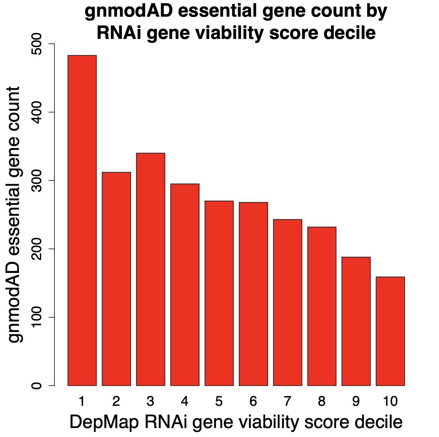
One of the major obstacles to eradicating this global health threat lies in the growing problem of insecticide resistance among its primary vectors, particularly the Anopheles mosquitos. These resilient insects have evolved to withstand the very agents intended to control them. In Anopheles gambiae, resistance to insecticides arises through a complex interplay of five distinct mechanisms: 1) behavioral shifts, such as alterations in host-seeking behavior; 2) cuticle thickening, which fortifies the mosquito’s exoskeleton; and at a molecular level, 3) alterations to target sites, 4) detoxification processes, and 5) insecticide binding.

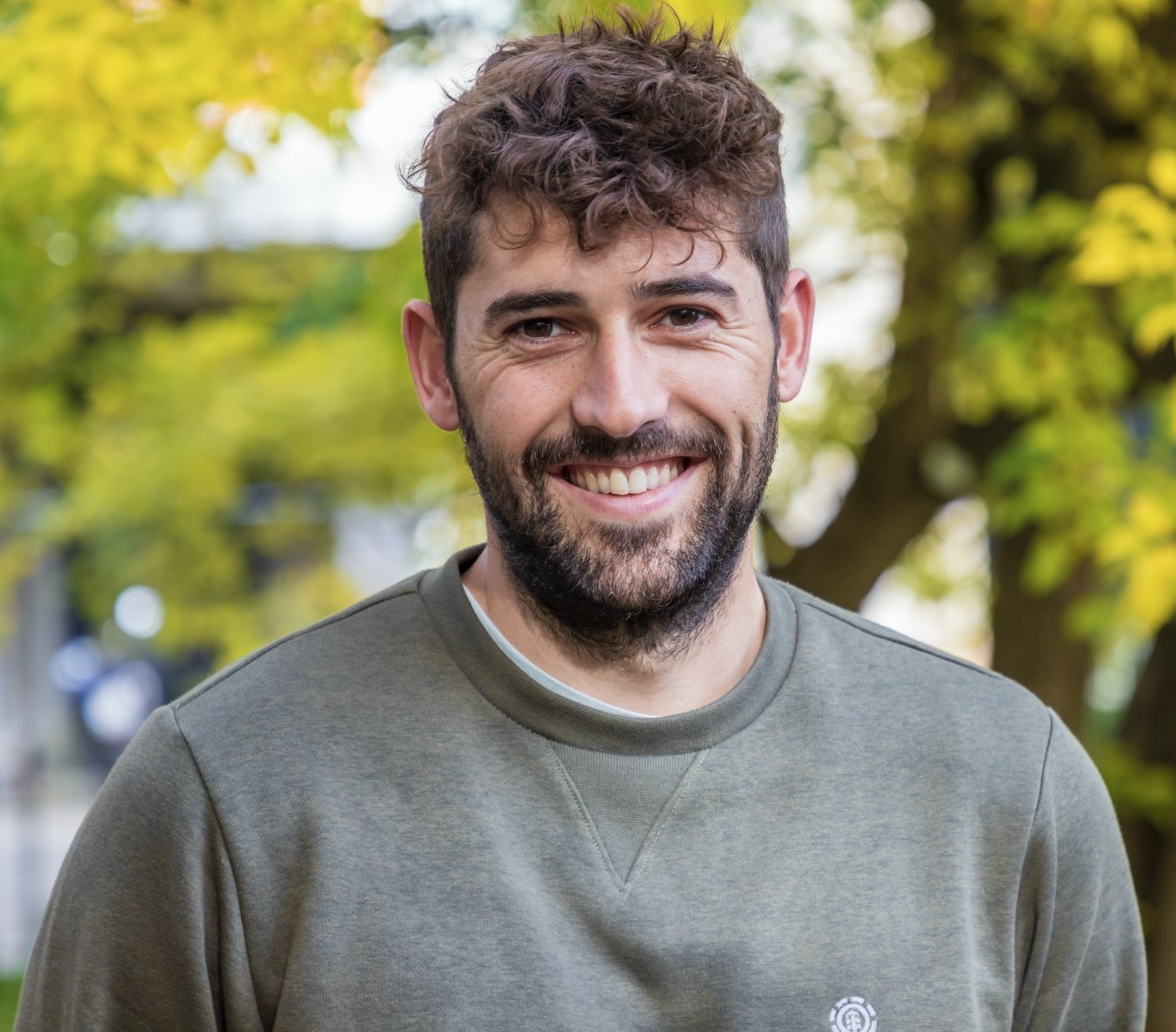
However, amidst these challenges, researchers like Patrick Hörner, a PhD student at Dr. Victoria Ingham’s lab in the Heidelberg University Hospital focus on investigating the impact of insecticide resistance phenotype on Plasmodium development in vivo.

Patrick Hörner, PhD student at the Heidelberg University Hospital

For his research, Patrick uses a combination of bioinformatics and molecular biology (e.g. RNAi) to identify the pathways and genes that influence vector competence and insecticide resistance status. To gain deeper insight into Patrick’s PhD research project, his motivations, the challenges he faces, and his aspirations in the global fight against malaria, we had the opportunity to interview him:

What initially sparked your interest in studying malaria and its vector mosquitos?

I have always been aware that malaria is a devastating disease and responsible for the death of many people, especially children in Africa. What triggered me to do my PhD in that field was actually a field trip to Namibia during my masters in 2019 though. I always had the urge to help people in some kind of way and I’m fascinated by the lifestyle of parasites, in particular in the interaction with their host species. So I did my master’s thesis project on the dog tapeworm Echinococcus granulosus at the University of Hohenheim and had the privilege to collect field samples in Namibia. When we collected our samples during the trip in this beautiful country, we were fortunate to get to know many very nice people in small villages in the so-called Caprivi strip in the north of the country. While we explained our research aims, most people only answered by asking in turn why we’re not researching malaria and they told us many stories about their encounters with the disease. You could really sense that malaria is one of the biggest threats they face.

Could you briefly tell us about your PhD research project?

My project deals with the problem of insecticide resistance of African malaria vectors and how we could overcome this major obstacle for the elimination of the disease. I’m particularly focusing on one major mechanism, that we think is related to resistance to insecticides and in the immune response against Plasmodium parasites. We try to find solutions on how we can manipulate mosquitos to either circumvent resistance or even tackle the malaria parasite inside the mosquito vector. The advantage of targeting the mosquito stages of Plasmodium has the advantage that they harbor the parasite’s life cycle stages where the lowest numbers are present, which is called the bottleneck.

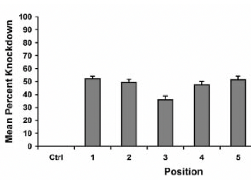
Some of the techniques you use are RNAi, what are some challenges or limitations you’ve encountered while working with RNAi in mosquitos?

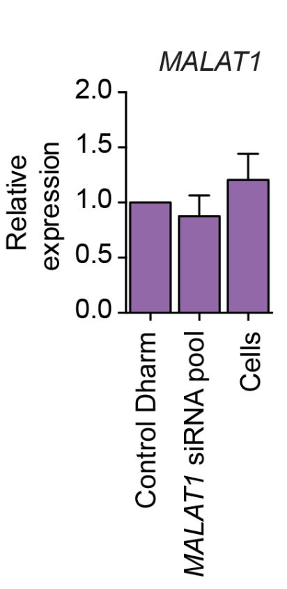
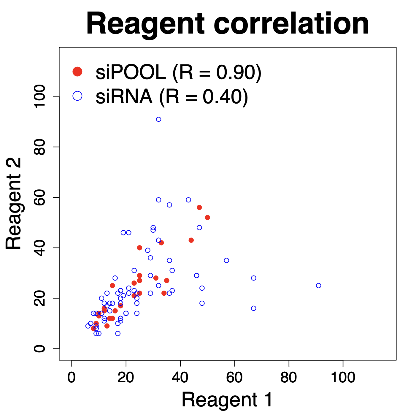
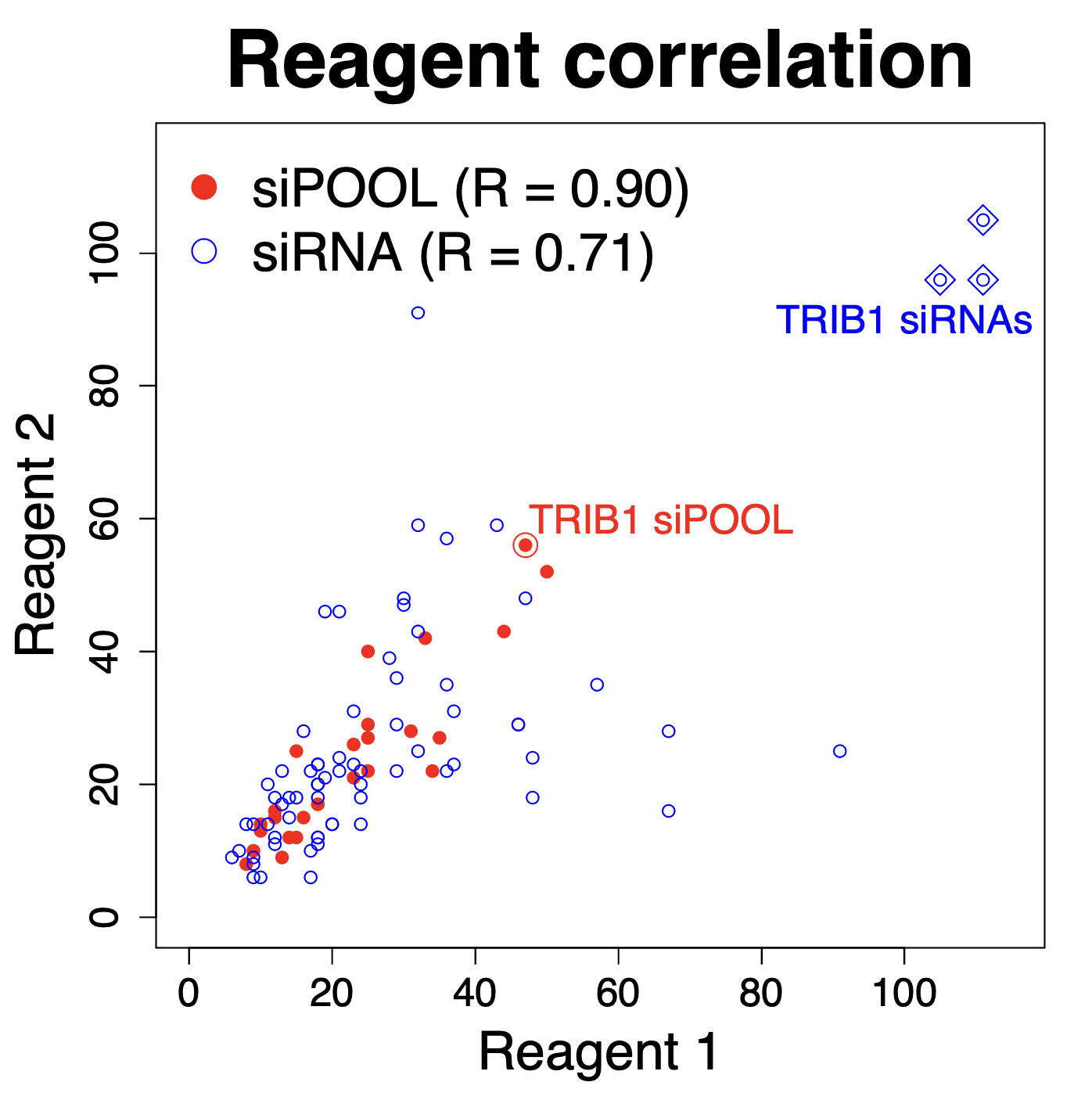
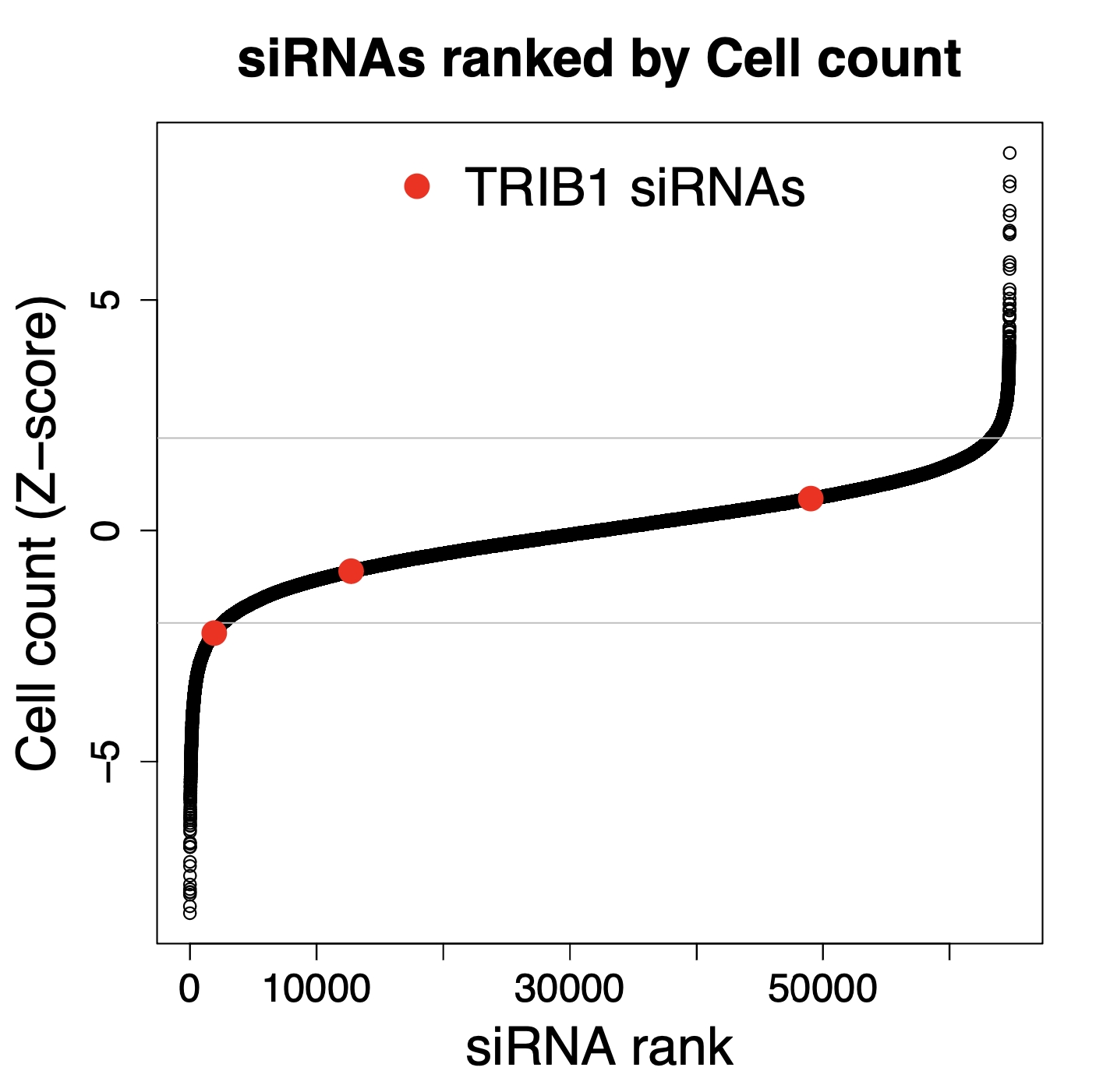
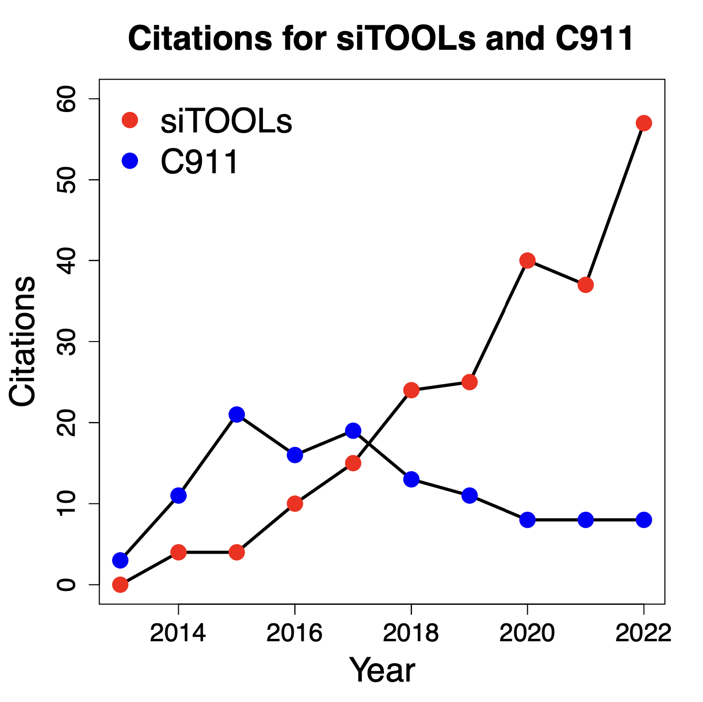
We are using RNAi to knock specific genes down in the mosquito by either injecting long dsRNAs or since the start of our collaboration with siTOOLs also siRNAs pools (siPOOLs). We look at how such knockdowns affect the development of the malaria parasites or the resistance status of the vector itself. Obviously, the first challenge is always to keep the mosquitos alive when you puncture them with a needle and inject the RNA into their thorax. That means you have to be very cautious and do a lot of practice sessions before you can actually do the experiments. The second obstacle is that you need a very high concentration of your RNA to effectively knock the genes down when you use “naked” RNA as we usually do, because a lot of it gets degraded before reaching the target.

How does your research on Anopheles gambiae tie into broader malaria control and prevention strategies?

Our research focuses on helping to improve vector control tools that are applied in field settings, like insecticide-treated bednets or new up-and-coming tools e.g. attractive targeted sugar baits. We also test the efficacy of currently used substances on such tools against mosquitos and parasites, especially the widely used pyrethroid insecticides. These insecticides have been causing widespread resistance in sub-Saharan Africa but are still applied on all insecticide-treated bednets, due to a lack of alternatives.

Are there any particular milestones or breakthroughs you’re aiming for in the near future within your research area?

I guess as a scientist you’re always aiming for breakthroughs but in the end, it’s very hard to define what that actually means. Probably there won’t be one specific breakthrough that eliminates malaria in the near future, as this parasite is very complex and adapts quickly. We’re always aiming to contribute to the pool of knowledge and tools in the fight against the disease because only the interplay of all existing measures, like vaccines, drugs, and vector control have a chance to keep the disease in check and eventually reach the common goal of eradication.

What precautions do you take when working with mosquitos?

When you’re doing experiments on uninfected mosquitos, we normally knock them down on ice first, so they can’t fly away. Of course, you get bitten here and there but that is just the nature of the work. It gets obviously trickier when you work with malaria-infected mosquitos. In this case, we have to keep them in a BSL-3 lab in humidified incubators and sort the ones we need for our experiments out, and kill them right away in a secured glass glove box. When you take those out of the BSL-3 you carry them in a sealed tube and inactivate the parasites at -80°C before you start your experiments.

If you would have not been a scientist, what other profession would you have chosen?

Although I’ve been playing and coaching soccer for my whole life, I’m an even bigger American football fan and an exchange semester to Penn State University during my bachelor’s only increased my love for the game. I would have always liked to go into coaching there, because of the high complexity of the game with endless individual and team tactics and techniques to explore for your team.

🦟🧬🦟🧬🦟

Working with mosquitos? We have reagents for RNA inteference (siPOOLs) as well as ribosomal RNA removal kits (riboPOOLs) for Aedes albopictus and Anopheles gambiae.  Request a quote here.

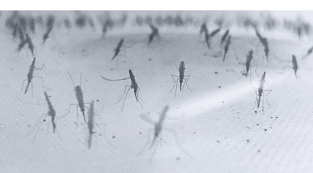
Image: Anopheles gambiae mosquitos (provided by Patrick Hörner).

Comparing silencing for CDS and 3′ UTR siRNAs

Comparing silencing for CDS and 3′ UTR siRNAs

CDS vs. 3′ UTR

In some cases, it may be preferable to only target the 3′ UTR of an mRNA.

For example, the CDS may be highly similar to a related paralog gene that should not be co-targeted.

Or a rescue experiment will be performed, and one would rather use the native CDS for rescue.

(Note that we can also provide rescue constructs for siPOOLs that target CDS. The rescue sequence uses the most common alternative codon in the targeted regions to ensure there is no silencing of the rescue construct.)

One common question is whether siRNAs against the 3′ UTR are as effective as those against the CDS?

Early results from 2004

An early paper by Hsieh et al. (2004) showed that siRNAs targeting the 3′ UTR (indicated as region 5 in the plot below) were as effective as those targeting CDS (indicated as regions 1-4):

siPOOL silencing

We have performed thousands of qPCR validation experiments on our siPOOLs and have also not found much difference between siPOOLs that target only CDS versus those that target only 3′ UTR.

The following plot shows the % remaining mRNA for siPOOLs that target only 3′ UTR (left) or only CDS (right):

And if we look at all possible numbers of CDS siRNAs, there is no observable trend. The horizontal line in the following plot shows the median siPOOL silencing (12.9% remaining RNA).

Design space

Given the focus on CDS for gene function, there may be an assumption that the 3′ UTR is relatively small and thus may not provide enough design space for siPOOL design.

The difference in CDS and 3′ UTR lengths for RefSeq coding genes is actually not that great:

The median CDS is ~1.3 kb, whereas the median 3′ UTR is ~1.0 kb. The mean is actually higher for 3′ UTR (~1.7 kb) than for CDS (~1.2 kb).

It should be noted, however, that there are some very short 3′ UTRs, and in those cases a 3′ UTR-only siPOOL will not be possible.

Conclusion

siPOOLs targeting only 3′ UTR should be as effective as ones only targeting CDS.

If our current design targets CDS, we can also do a redesign that only targets 3′ UTR.

Complex siRNA pooling is especially important for silencing lncRNAs

Complex siRNA pooling is especially important for silencing lncRNAs

One advantage of pooling siRNAs is that the pool tends to silence about as well as any single siRNA. That is why independent complex siRNA pools (siPOOLs) for the same gene have much more similar and better average silencing than single siRNAs or mini-pools (Dharmacon), as discussed in our last blog post.

Another advantage of complex siRNA pools is that you can cover more regions of the target gene. This may be especially important when silencing lncRNAs, which can be very long and whose structure may cause certain regions of the transcript to be inaccessible to RISC.

MALAT1

A good exemplar of this phenomenon is MALAT1, whose transcripts are nearly 9 kb and whose secondary and tertiary structure is important for its cellular function.

Two groups who used single siRNAs or mini-pools (Dharmacon) found poor silencing for MALAT1.

Stojic et al. (2018) used a Lincode mini-pool (Dharmacon) and saw almost no silencing:

That was despite using a very high siRNA concentration (50 nM) in a cell line where RNAi normally works very well (HeLa).

Lennox and Behlke (2016) used several single siRNAs at two concentrations (1 nM and 10 nM), again in HeLa cells. They found highly variable silencing, where most siRNAs had poor silencing, and a small number worked fairly well at the higher concentration (10 nM):

Nuclear IncRNA: MALAT1

If one were to randomly choose an siRNA, there is only a 25% chance (3 / 12) that it would give decent silencing at 10 nM (using 30% remaining RNA as cutoff for decent silencing). And none would be considered decent at 1 nM.

Of note, this paper is often cited as a reason for not using RNAi for lncRNAs (the authors, from IDT, recommend using ASOs).

The results from Stojic et al. were quite poor (the Lincode mini-pool hardly silenced), and could be due to reagent quality.

siPOOLs effectively silence MALAT1

The results from Lennox and Behlke are more in line with what we’ve observed when researching the silencing of individual siRNAs versus complex siRNA pools (siPOOLs). Some siRNAs are much better than others. And, as mentioned earlier, a complex pool of siRNAs tends to silence like the best constituent siRNAs.

Given the best-silencing-siRNA selection from complex siRNA pooling and the increased transcript coverage, we would expect siPOOLs to give better silencing than what these authors found.

Our 2 independent siPOOLs for MALAT1 give silencing of 16.5% and 24.4% remaining RNA, respectively, at 1 nM:

MALAT1 Silencing by siPOOLs

If we compare our reagents to those used by Lennox and Behlke, we see that there is much broader transcript coverage. As mentioned, this could be especially important for lncRNAs like MALAT1 that have extensive secondary and tertiary structure.

Lennox and Behlke siRNAs (used individually):

siPOOL #1 siRNAs (used together):

Conclusion

Two factors make complex siRNA pools (siPOOLs) especially well suited for silencing lncRNAs:

  1. Complex siRNA pools tend to silence like their best constituent siRNAs. A single siPOOL silences about as well as the best single siRNA. For targets with high silencing variability, siPOOLs are far superior to single siRNAs or mini-pools (Dharmacon).
  2. By using a complex siRNA pool, more of the gene can be covered. For targets with lots of secondary and tertiary structure, siPOOLs give you the best chance of targeting an accessible region.
siPOOLs: robust reagents for gene silencing

siPOOLs: robust reagents for gene silencing

Although we talk a lot about off-targets, one of the main advantages of siPOOLs (complex siRNA pools) compared to single siRNAs or mini-pools (Dharmacon) is that they provide near optimum silencing of target genes. Two siPOOLs for the same gene give very similar knock down levels, and their silencing is around the best of any single siRNA. Given how many candidate siRNAs there are for a gene, and how difficult it is to accurately predict silencing levels, this makes siPOOLs the best choice for gene silencing.

The following plot, comparing independent siPOOLs and siRNAs for the same target gene, shows that siPOOLs for the same gene give more similar silencing than do siRNAs (these are Ambion Silencer Select siRNAs).

We see that the correlation for independent siPOOLs is nearly twice that for independent siRNAs.

(Note that for siRNAs we are doing all pairwise comparisons for 3 siRNAs per target gene. Randomly selecting 2 siRNAs per gene gives similar R values.)

In the above plot, we removed 3 siRNAs that did not work, for the gene TRIB1. TRIB1 has some association with the nucleus and has a short mRNA half life, both of which are factors associated with poor gene silencing.

The following plot shows the TRIB1 siPOOLs and siRNAs.

Note that including these non-functional siRNAs actually improves the reagent correlation, though not for a good reason!

We also see that independent TRIB1 siPOOLs give very similar silencing and it’s much better than for the siRNAs. In our experience, if a siPOOL does not work well for a gene, designing a second siPOOL does not substantially improve things, as the poor silencing is normally a feature of the target gene itself. ~50% silencing is probably about the best one can expect for this gene.

Just because siRNAs do not give any on-target silencing, this does not mean they can’t show up as hits in screening assays. Because most of the downregulation is in off-target genes (due to the seed effect), each of those TRIB1 siRNAs may silence nearly 100 genes.

We looked at a genome-wide RNAi screen that included these 3 Silencer Select siRNAs. We see that one of them gives a fairly strong phenotype (Z-score < -2 for cell count), even though the siRNAs do not silence their on-target gene.

Screening with siPOOLs is the smarter alternative, as you can be confident that they provide near optimal on-target silencing and have less off-target effects.

Cutting the Gordian Knot of RNAi off-targets

Cutting the Gordian Knot of RNAi off-targets

The C911 siRNA control generated a lot of excitement in the RNAi world when it emerged ~11 years ago. A former colleague, who was a pioneer in the commercialisation of RNAi, described it then as the biggest breakthrough in the last 10 years of RNAi research.

The idea of the C911 control is to get rid of the on-target effect of the siRNA by using the complement of bases 9-11, while retaining any off-target (seed-based) effects of the siRNA, which are mostly dictated by the bases in positions 2-8.

If the observed phenotype of the siRNA is due to an off-target effect (rather than silencing of the on-target gene), the C911 version will show the same phenotype. i.e., because it is not silencing the target gene, the phenotype must come from an off-target effect.

Despite the initial excitement, the C911 approach did not become that widely used. There are a number of drawbacks to the strategy, perhaps foremost being that new reagents must be ordered and the assay set up to run again. We’ve compared the validation of low-complexity RNAi reagents to the old lady who swallowed a fly.

The best strategy is to avoid getting entangled in off-targets in the first place. And that seems to be the approach preferred by the research community.

The following plot shows Google Scholar citations for siTOOLs (i.e., papers using our reagents) and the C911 method paper.

We see that after an initial adoption period, use of C911s tapered off and it has levelled out in recent years.

None of this suggests that C911s are bad. For single siRNAs or Dharmacon pools, they are indeed an effective control. But the inconvenience of the method has probably hindered its adoption.

The convenience and robustness of the siPOOL are its great advantages. The siPOOL approach ensures maximum on-target silencing and a minimum of off-target effects. We look forward to supporting more great research in the coming years.

Our Team’s Favorite RNA Molecules🧬

Our Team’s Favorite RNA Molecules🧬

At siTOOLs we are celebrating RNA day (Aug. 1st) the entire month with a focus on all things RNA and a promotion on siPOOLs and riboPOOLs.

RNA day is celebrated the 1st of August, since the RNA codon that initiates protein synthesis is made of the following nucleotides: adenine (A), uracil (U), and guanine (G). AUG codes for the amino acid methionine (Met) in eukaryotes and formyl methionine (fMet) in prokaryotes.

RNA is one of the most versatile biomolecules in existence and the focus of siTOOLs research, so we asked the siTOOLs team which is their favorite RNA molecule.✨

1. Dr. Michael Hannus – Founder and Managing Director:

2. Dr. Nicola Conci – RNA-Seq Specialist & Bioinformatician:

3. Matthias Weiss- Head of Production:

4. Kevin Wobedo – Scientific Sales & Marketing:

5. Dr. Laura Leiva – Scientific Marketing:

6. Jessie Midgley – Junior Sales Manager:

RNAi vs CRISPR: RNAi even better at finding essential genes

RNAi vs CRISPR: RNAi even better at finding essential genes

Which technology is better, RNAi or CRISPR?

The best answer to this question, like so many others is, it depends.

If cells can adapt and compensate for loss of the gene, or you want to titrate gene levels (important in drug discovery), then RNAi will be better.

If a gene’s transcripts have lots of secondary structure and must be silenced to 99.9% in order to see an assay phenotype, then CRISPR may be better.

We have used two large datasets to attempt to answer the following question: is RNAi or CRISPR better at identifying essential genes?

The first dataset is the BROAD Institute’s Dependency Map (DepMap). It has both RNAi (shRNA) and CRISPR (Cas9) screens from over 700 human cell lines, using hundreds of thousands of reagents. Both types of reagents were used to do pooled screening for cell viability.

The second dataset, also from the BROAD Institute, is called gnomAD. It has genome and exome sequencing for over 100K humans. Based on how frequently mutations are found in the sequenced genomes/exomes (and what type of mutations are preferred), an essentiality score can be assigned to every human gene. It’s the ultimate test (within ethical limits) of whether a gene is essential to humans.

Our approach was as follows:

  • for each gene, get the median DepMap viability score across the 700+ cell lines
    • done separately for RNAi and CRISPR screens
  • for each gene, retrieve the gnomdAD pLI score (probability that loss-of-function not tolerated)
    • higher values means the gene is considered more essential
    • genes with pLI > 0.9 are classified by gnomAD as essential

If we look at the top 200 genes in each of the RNAi and CRISPR datasets (note: 70 genes are common to both lists), we see that the top 200 genes from RNAi screening are more essential (as measured by pLI) than are the top 200 genes from CRISPR screening. (note that the curves show the running mean for 30 genes)

Eventually the curves do converge, but for the top genes, we see that those found by RNAi are more essential.

Alternatively, if we group the genes from the CRISPR and RNAi screens into deciles for cell viability score, we again see that the results from the RNAi screens are more consistent with gnomAD.

In the following plots, we look at the number of gnomAD essential genes (defined as pLI > 0.9) in each of the deciles. Decile 1 has the top 10% of genes for reducing cell viability (most essential), whereas Decile 10 has the bottom 10% (least essential).

For CRISPR screens, we see that the top 2 deciles show markedly more gnomAD essential genes. But after that, the counts flatten out. There is little difference in the number of gnomAD essential genes in deciles 3 through 10.

The results from RNAi screening show a fairly steady decline in gnomAD essential genes in deciles 1 through 10. Which is what one would expect. Genes that increase cell count should tend to be less essential. i.e., decile 10 should have the smallest number of essential genes. That is what we see with the RNAi screens, but not with the CRISPR screens.

Conclusion

RNAi and CRISPR screens can both pull out genes found to be essential in the gnomAD dataset.

However, the top genes from RNAi screening tend to be a bit more essential in real-life experiments (i.e., the humans from the gnomAD dataset).

Furthermore, the trend for gnomAD essential gene counts through the ranked datasets makes more sense for RNAi screens than for CRISPR screens.

CRISPR may be a newer technology, but that does not necessarily make it better than RNAi.

Both have their advantages and disadvantages, which we will discuss more in future blog posts.

It should also be noted that two of the main disadvantages of RNAi screening (seed-based off-target effects, and variability in silencing between different siRNAs) have been addressed by siPOOLs.

In an upcoming blog post, we will take a closer look at genes that gave different results in the DepMap RNAi and CRISPR screens.

Chemical modifications only shift the siRNA seed profile

Chemical modifications only shift the siRNA seed profile

In the last post, we saw that chemically modified ON-TARGETplus siRNAs still have a strong seed effect.

The seed-based off-target effects (measured by correlation of reagents with the same 7mer seed) were as strong for chemically modified ON-TARGETplus (R = 0.50) and Silencer Select (R = 0.59) as what we typically see with unmodified siRNAs (Qiagen, siGENOME, or Silencer).

Chemical modification must not prevent seed-based target recognition, because RISC uses the seed to scan the transcriptome for target sites. Because of how RISC presents the guide strand seed region for target scanning, the binding energy for finding an on-target site (19-base complementarity) versus an off-target site (6/7-base complementarity) is nearly the same. It’s not like a microarray oligo, where more extensive complementarity leads to stronger binding. The seed is driving this site recognition, so any modification that eliminates its binding will make the siRNA ineffective.

The chemical modifications added by Ambion and Dharmacon do not prevent seed binding, but instead change the efficiency of different bases at certain positions, in effect changing the seed profile of off-target sites.

The following heatmap shows the cell viability scores from 9 genome-wide siRNA screens. The average viability score for all siRNAs with a specific base at a specific position was calculated (shown are guide positions 1-9). If the value is red, it means siRNAs with the base at that position tend to be more lethal.

The first 4 columns are from screens using chemically modified, Silencer Select siRNAs (S+). The next 2 columns are from screens using unmodified, Silencer siRNAs (S). And the last 3 are from screens using unmodified, Qiagen siRNAs (Q).

We see that for some bases (e.g. 2C, top row), siRNAs tend to be non-toxic regardless of whether or not they are chemically modified (S+, S, and Q all show deep blue).

But there are other positions where the chemically modified siRNAs are very different from the unmodified siRNAs.

For example, the bottom row shows that 6G tends to be very toxic in unmodified siRNAs, but is not toxic in Silencer Select (chemically modified) siRNAs. On the other hand, 6U (towards the middle row) looks to be toxic for Silencer Select siRNAs but have the opposite effect for unmodified siRNAs.

Whatever the chemical modification for Silencer Select is (has not been made public), it appears to make seed off-targets stronger when position 6 is a U, and weaker when position 6 is a G.

If we compare the effect on cell viability of Silencer Select vs ON-TARGETplus siRNAs from the Tan and Martin screen (subject of last post), we also see strong differences in the effect of having a U or a G at position 6.

The following plot shows the toxicity rank of seed bases in Silencer Select siRNAs vs ON-TARGETplus siRNAs. Bases towards the origin (e.g., 2C) tend to make siRNAs non-toxic for both types, whereas bases towards the top right (e.g., 2G) tend make make siRNAs toxic for both types. Bases that fall off the diagonal tend to be toxic for one type and non-toxic for the other.

We see that 6U is toxic for Silencer Select siRNAs (as also seen in the heat map) and ON-TARGETplus siRNAs, like the unmodified siRNAs from the heat map, tend to be non-toxic. And the effect is similar to the heat map for 6G: toxic for Silencer Select and non-toxic for ON-TARGETplus (and unmodified in heat map).

Conclusion

Chemical modification does not get rid of seed effects, as evidenced by the strong phenotypic correlation of modified siRNAs with the same seed sequence. Rather, modifications tend to change the effectiveness for specific bases in eliciting seed-based silencing.

One suggestion would be to design a chemically modified siRNA library that avoids bases that tend to be toxic (e.g., 6U for Silencer Select).

However, there are a few problems:

  • The heat maps and scatterplot only show tendencies. There is still variation within those positions. While 2G tends to be non-toxic for Silencer Select, there are still lots of toxic siRNAs with that sequence.
  • Bases that reduce toxicity may be doing so because they tend to reduce target recognition. For example, 2C is also associated with poorer on-target silencing. Using only 2C for siRNAs could thus result in a library that is not as efficient at on-target silencing.
  • Finally, these suppliers have already produced their siRNA libraries. i.e., those bases have already been used.

The only reliable way to both reduce the off-target effect (via dilution of seeds) and maintain robust on-target silencing is by using siRNA pools (siPOOLs).

ON-TARGETplus siRNAs have strong off-target effects (despite chemical modification)

ON-TARGETplus siRNAs have strong off-target effects (despite chemical modification)

History of chemical modifications

Chemical modification has long been proposed as a way to limit the off-target effects of siRNAs.

The earliest siRNAs from the two main commercial suppliers (siGENOME from Dharmacon/Horizon Discovery, and Silencer from Ambion/ThermoFisher) were quickly replaced with new chemically-modified siRNAs (ON-TARGETplus from Dharmacon, and Silencer Select from Ambion).

We have already seen that Silencer Select siRNAs, despite their chemical modification, maintain a strong off-target seed effect.

The phenotypic correlation between siGENOME (unmodified) and ON-TARGETplus (chemically modified) low-complexity (4-siRNA) pools for the same gene was shown to be very poor.

However, showing a direct seed effect of ON-TARGETplus siRNAs using published data is not straightforward, since Dharmacon (unlike Ambion) has not made their siRNA sequences publicly available.

Here, for the first time, we show massive seed-based off-target effects from ON-TARGETplus siRNAs.

Seed off-target effects from ON-TARGETplus siRNAs

Tan and Martin (2016) provide a dataset that includes 4 different ON-TARGETplus siRNAs for nearly 700 genes, screened for their effect on nuclear area.

We were also able to find a paper that provides sequences for ON-TARGETplus siRNAs. Those sequences were assigned to the siRNAs from the Tan and Martin screen (details on sequence assignment provided at end of post).

The intraclass correlation (ICC) is a measure of reproducibility of measures of the same group, e.g. siRNAs with the same target gene, or siRNAs with the same 7mer seed.

The ICC for ON-TARGETplus siRNAs with the same gene was only 0.09.

However, the ICC for ON-TARGETplus siRNAs with the same 7mer seed was much higher: 0.50.

Despite chemical-modification, the phenotype of ON-TARGETplus siRNAs is still mostly driven by off-target seed effects.

To show these ICCs graphically, here is a plot with pairs of siRNAs for the same target gene (2 of 4 siRNAs chosen randomly for each gene). [ note that some outliers were removed to assist comparison with same-seed siRNAs]

And here is the plot with pairs of siRNAs with the same 7mer seed:

Conclusion

Chemical modification does not get rid of seed-based off-target effects.

The only effective way to robustly eliminate these effects is with high-complexity (30+ siRNA) pools (siPOOLs).

Technical notes

In order to determine the sequence of the ON-TARGETplus siRNAs from the Tan and Martin screen, the sequences from the supplementary materials of Kim et al. were assigned in order to the siRNAs sorted by catalog number. It is possible that some of the sequences thus assigned were not correct (e.g. Tan and Martin may have used different siRNAs from those listed in Kim et al. for some of the genes), in which case the observed seed effect is actually underestimated.

A journey into the gut microbial control center: small RNA’s influence on Bacteroides thetaiotaomicron’s metabolism

A journey into the gut microbial control center: small RNA’s influence on Bacteroides thetaiotaomicron’s metabolism

The gut model organism Bacteroides thetaiotaomicron

Bacteroides thetaiotaomicron is a commensal bacterium that inhabits primarily the human large intestine and is considered one of the most important members of this microbial community. B. thetaiotaomicron is a highly versatile microbe, capable of utilizing a wide range of carbohydrates including those that are indigestible by human enzymes. It breaks down complex polysaccharides from plant cell walls and other dietary sources, producing short-chain fatty acids (SCFAs) that are an important energy source for humans. Furthermore, it has also been shown to play a crucial role in our immune system development, through the production of regulatory T-cells that help prevent autoimmune disorders.

It’s no wonder Bacteroides thetaiotaomicron has been chosen as a model representative of the gut microbiota. B. thetaiotaomicron is widely spread in human populations and relatively easy to grow and study under laboratory conditions. In a study by Ryan et al. (2020), differential RNA sequencing (dRNA-Seq) was used to generate a single-nucleotide resolution transcriptome map of B. thetaiotaomicron. High-resolution RNA-sequencing served as a tool to explore the role of small RNA molecules in regulating metabolism in the gut bacterium Bacteroides thetaiotaomicron.

By comparing different laboratory growth conditions, the researchers identified various small RNA molecules that exhibited differential expression patterns. These small RNAs were found to be associated with the regulation of key metabolic pathways in the bacterium. The authors suggest that these findings could have implications for understanding the interactions between gut microbes and the host and for the development of new therapies for metabolic diseases. Overall, the study highlights the importance of transcriptome mapping in uncovering novel regulatory mechanisms in bacterial metabolism. Furthermore, the results shed light on the intricate regulatory networks within this gut microbe and provide insights into its adaptation to different nutrient environments.

Note: Our Pan-Prokaryote riboPOOL played a small but significant role in this study. Prior to RNA sequencing, the Pan-Prokaryote riboPOOLs kit was used for ribosomal RNA depletion. Our Pan-riboPOOLs are a versatile solution that allows for simple mono- and multitranscriptomic studies using a single-step rRNA depletion for a phylogenetic group (e.g., bacteria, fungi, or mammals).

A Brief Interview with Dr. Daniel Ryan

Dr. Daniel Ryan, postdoc at the Helmholtz Institute for RNA-based Infection Research

We interviewed the first author of the study: “A high-resolution transcriptome map identifies small RNA regulation of metabolism in the gut microbe Bacteroides thetaiotaomicron”. Dr. Daniel Ryan is a postdoc at the Helmholtz Institute for RNA-based Infection Research located in Würzburg, Germany. He is a member of the Westermann Lab and his research focuses on non-coding RNAs and RNA-binding proteins in the human gut commensal Bacteroides thetaiotaomicron.

He further explained the process of generating a high-resolution transcriptome for Bacteroides thetaiotaomicron and the exciting parts of being an RNA research scientist.

  1. Can you briefly explain the main findings of your research and what motivated you to study small RNA regulation in Bacteroides thetaiotaomicron?

The gut microbiota has recently attracted significant attention from the scientific community due to its impact on human health and physiology. Various diseases, including inflammatory bowel disease (IBD), diabetes, colon cancer, and depression, have been linked to an imbalanced microbiota, also known as dysbiosis. Furthermore, a healthy gut microbiota plays a crucial role in preventing invasive pathogens from gaining a foothold and establishing infections. My research at the Westermann Lab aims to understand the diverse interactions between gut microbes and their host. To achieve this goal, we utilize the gut model organism Bacteroides thetaiotaomicron (B. theta), an anaerobic, non-spore forming predominant member of the healthy gut microbiota.

My foray into small RNA biology started during my Master’s thesis work at the KU Leuven, Belgium where I investigated the regulatory networks of sRNAs in E. coli. I came to appreciate the immense regulatory potential of these non-coding molecules in governing rapid responses to diverse environmental cues. A few years later, during my PhD research at KIIT University, India, I had the opportunity to delve into the roles of sRNAs in regulating acid stress survival and virulence programs in Salmonella, the pathogen responsible for causing typhoid. Having gained extensive experience in studying sRNAs and their intricate regulatory networks, I shifted gears to the gut microbiota as the focus of my postdoctoral research. After more than a decade of involvement in the field of sRNAs, I remain highly enthusiastic about uncovering novel sRNAs and investigating their intricate interactions within diverse organisms. Ultimately, my aim is to reveal regulatory cascades and pathways that can be harnessed to improve human health.

  1. What tools were necessary to create a high-resolution transcriptome map for Bacteroides thetaiotaomicron, and what challenges did you encounter during this process?

In order to construct a high-resolution transcriptome map of B. theta, it is crucial to extract RNA of high quality from representative and diverse conditions that effectively stimulate gene expression. This is easier said than done, since one of the main challenges of working with gut microbes is their anaerobic nature and often cumbersome culture conditions. To ensure optimal anaerobic conditions, all media and equipment used for bacterial culture must be degassed to remove oxygen, which can be toxic and inhibit growth. Once robust and reproducible growth can be achieved, RNA is extracted, sequenced and analyzed to obtain a single nucleotide resolution of the transcriptome. I then employed a suite of bio-informatics tools to annotate the transcriptome and subsequently manually validate and edit each feature. Although this final step is time-consuming and labor-intensive, it is essential for obtaining a high-quality and reliable result.

  1. Were there any unexpected or surprising findings in your research?

I was delighted to discover that the number of potential sRNAs in B. theta was similar to that of other model organisms, such as E. coli and Salmonella. Moreover, the vast majority of these sRNAs had no known homologs in these well-known species. This suggests that B. theta has undergone functional adaptations specific to its niche, which is primarily the human large intestine. Consequently, I anticipate a wealth of novel biological insights, potentially revealing new modes of interaction and target regulation.

  1. Are there any potential applications or implications of your research for human health, such as developing targeted therapies or interventions for gut-related disorders?

In order to develop effective targeted therapies, it is crucial to first and foremost “know your target”. Going in blind is never a good strategy and this is where I see the potential of this work. With this high-resolution transcriptome and the “Theta-Base” browser, we have provided a framework to discover and identify novel genes and sRNAs that can further be investigated as potential targets to regulate or modulate activity. These newly identified targets whether coding or non-coding can be exploited to modulate B. theta to achieve specific functions for instance, they could be used to exclusively metabolize a particular carbon source or prevent the consumption of a specific metabolite. While this example is simplistic, several laboratories are already conducting pilot studies in this area, offering promise for the future of targeted medicine.

  1. What would you say are some of the challenges or gaps in knowledge that need to be addressed in the field of gut microbiota research?

One of the overarching challenges in gut microbiota research is distinguishing between correlative and causative effects. It is therefore imperative to develop protocols and methodologies that delve into various phenomena at a detailed level before drawing reliable conclusions.

There are also specific challenges to address, particularly regarding the creation of microbial consortia that accurately reflect the composition of the gut microbiota.  This is not easy considering the vast numbers of bacteria and their complex interactomes that make up a healthy microbiota. Moreover, models representing the human intestinal niche, which harbor these diverse microbial communities, need further refinement to better reflect this complex environment.

  1. Finally, what is your favorite part of being an RNA research scientist?

As an RNA research scientist, what I find most fascinating is the varied range of roles that this molecule can play. From intricate structural scaffolds to subtle enzymatic and regulatory functions, RNA displays a multitude of capabilities, and witnessing these firsthand is truly captivating.

Biocabulary

Differential RNA sequencing (dRNA-Seq) is a technique used to identify transcriptome features and define overall transcriptomic architecture, such as transcription start sites, terminators, non-coding RNAs, coding RNAs, promoters, etc.

A transcriptome map is a comprehensive profile or catalog of all the RNA molecules (transcripts) produced by an organism or a specific cell type under particular conditions. It provides a snapshot of the active genes and their expression levels within the cells or tissues being studied.

error

Like what you see? Mouse over icons to Follow / Share